Auditory Ossicles (Ear Bones)
The human ear is composed of three parts – the external/outer ear, middle ear, and internal/inner ear. The middle ear region holds the three smallest bones of the body, collectively known as auditory ossicles.
There are three bones in each ear, so there are a total of 6 auditory ossicles in the body. These are the first bones to ossify and be fully mature at birth, so they do not grow anymore.
Where are the Ossicles Located in the Ear
The middle ear has a hollow space called the tympanic cavity, an air-filled cavity in the temporal bone’s tympanic part. This is where the three ear ossicles are located.
Structure and Anatomy of the Bones in the Middle Ear
1. Malleus: It is the outermost of the three bones, shaped like a hammer, hence the name (malleus is the Italian for hammer). It is structurally divided into several parts – the head, neck, spatulate, lateral, and anterior processes, and the manubrium (handle) of the malleus. The bone stays connected to the tympanic membrane or eardrum through the manubrium.
Three ligaments hold this bone in place as it stays suspended in the anterior part of the middle ear.
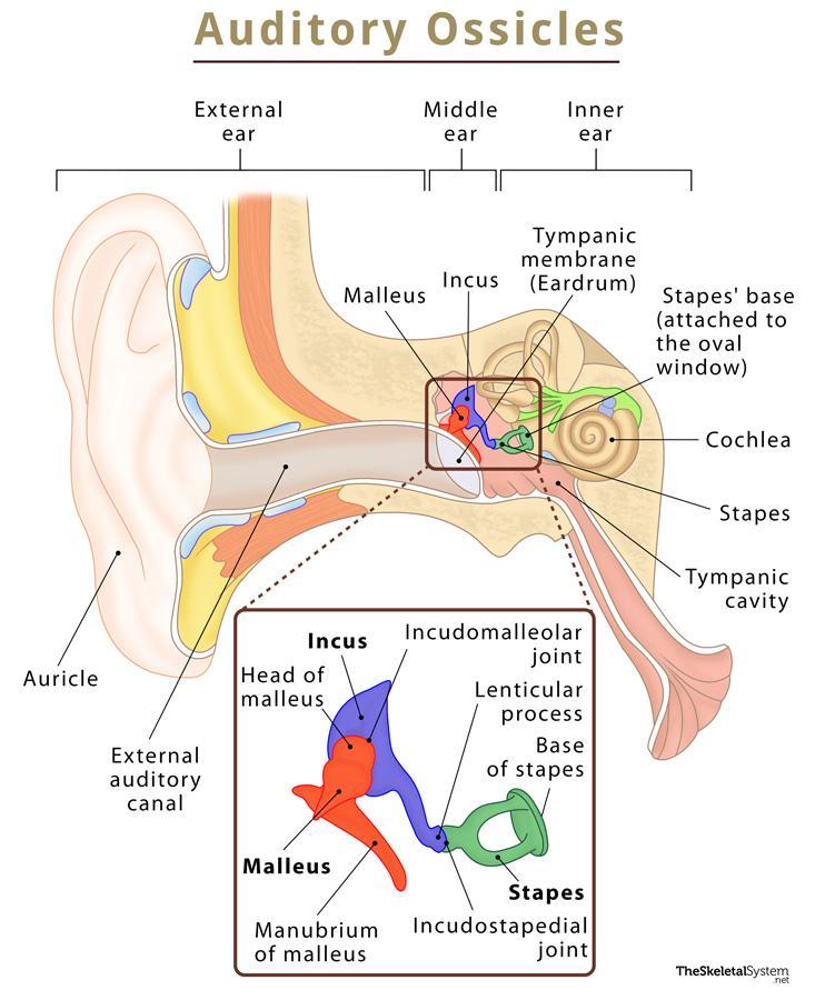
2. Incus: The incus or anvil is an anvil-shaped bone located after the malleus, consisting of a body and long and short limbs. It is also held in place by ligaments, forming two synovial joints with the other two ear bones.
3. Stapes: The smallest of the three bones, it is located in the innermost part of the middle ear. This stirrup-shaped bone has four parts – The head, base, and the anterior and posterior limbs.
Its head articulates with the incus, while its base or footplate lies in the oval window of the tympanic cavity, connecting the middle ear with the inner ear. This point of connection is known as tympanostapedial syndesmosis.
Articulations
- Incudomalleolar Joint: Synovial joint between the malleus (head) and incus (body).
- Incudostapedial Joint: Synovial joint between the incus (lenticular process of long limb) and the stapes (head).
Muscles
Two muscles are associated with the middle ear:
- Tensor tympani
- Stapedius muscle
Functions of the Auditory Ossicles in Hearing
The sound waves first make contact with the external auditory canal and vibrate the eardrum, which in turn moves the ear ossicles one after the other – taking the sound waves to the inner ear through the oval window of the tympanic cavity. So, the primary function of these tiny bones is to transport sound vibrations from the external ear to the inner ear.
At the tympanostapedial syndesmosis, the footplate of stapes vibrates and leads to the movement of the fluid in the cochlea in the inner ear. The waves are then converted to electrical impulses that are picked up by the receptor cells within the fluid and transmitted to the brain through the auditory nerves. Once the brain translates these impulses as sound, we finally hear them.
FAQs
Ans. Since the malleus is located closest to the external ear, it is the first of the three ossicles to vibrate, followed by the incus through the incudomalleolar joint. Lastly, the vibrations reach the stapes through the incudostapedial joint and are transmitted to the inner ear.
References
- Auditory Ossicles – Kenhub.com
- Anatomy, Head and Neck, Ear Ossicles – Ncbi.nlm.nih.gov
- Bones of the Ear – Innerbody.com
- Anatomy and Physiology of the Ear – Stanfordchildrens.org
- Slide Show: How You Hear – Mayoclinic.org