Malleus
Table of Contents:
Published on November 14th 2022 by staff
What is the Malleus
The malleus is the largest of the three middle ear ossicles. The name derives from the Latin word for a mallet or hammer.
Where is the Malleus Located
It is the most lateral of all the middle ear ossicles, which means it is the first ear bone found when entering the middle ear from the external ear. It is directly attached to the tympanic membrane or eardrum.
Quick Facts
| Type | Irregular bone |
| Size (in a typical adult) | Height: About 8 mmWidth: About 3 mm |
| How many are there in the human body | 2 |
| Articulates with | Incus |
Functions
It is the first ear ossicle to move when a sound vibration reaches the tympanic membrane. So, its primary function is to transfer these sound vibrations from the tympanic membrane to the incus so it can be sent on to the inner ear through the stapes and oval window. This is how sound makes its way to our brain for us to hear and make sense of it
Anatomy
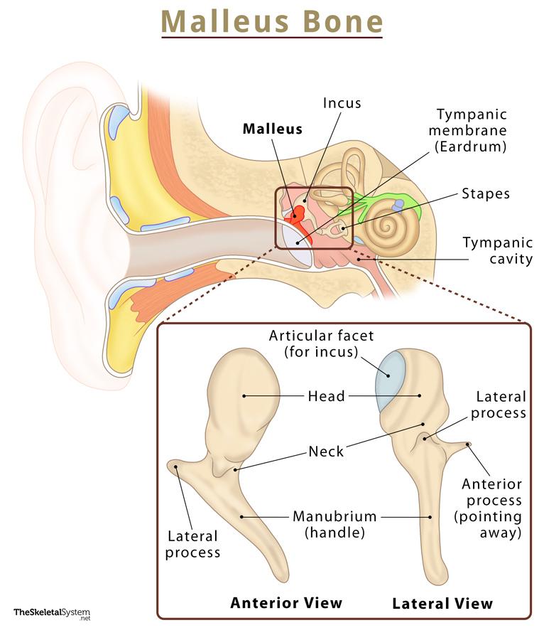
As mentioned above, this bone resembles a hammer in shape, having a head, neck, and multiple essential processes and landmarks.
Head: It is the oval, saddle-shaped posterior surface of the bone that articulates with the incus, forming the incudomalleolar joint, which is a synovial joint.
Neck: It is the narrow region just below the head, lying on top of the flaccid portion of the tympanic membrane, the pars flaccida. Inferior to it is a bony prominence where the malleus’s processes attach.
Manubrium (Handle): This is the most notable landmark of the malleus, extending downwards as it curves and narrows towards its tip. The manubrium’s lateral wall connects with the tympanic membrane. The small projection on its medial wall serves as the point of insertion for the tensor tympani muscle.
Lateral process: It is the small cone-shaped projection at the base of the manubrium. The anterior and posterior malleolar folds hold this lateral projection in its place where it attaches to the upper part of the eardrum.
Anterior process (Rau’s/Folian process): Another cone-shaped projection, it is much longer and more pronounced than the lateral process. It is located between the malleus’s neck and the lateral process, attaching to the middle ear at the anterior wall.
Ligament Attachments
Like the other two ear bones, the malleus is suspended in place by only the following three suspensory ligaments:
- Anterior malleal ligament (Casserio’s ligament): Attaches the malleus’s neck to the tympanic cavity’s anterior wall.
- Superior malleal ligament: Attaches the head to the tympanic cavity roof.
- Lateral malleal ligament: Attaches the malleal head (or neck) to the back of the tympanic notch.
References
- Malleus: RadioPaedia.org
- Malleus: IMAIOS.com
- Auditory ossicles: KenHub.com
- Malleus, Incus, & Stapes: Study.com
- Malleus: HealthLine.com