Skull Bones
The skull is one of the most vital bony structures of the human body, as it houses and protects the most important organs, including the brain. There are 29 bones (including the hyoid and middle ear bones) that comprise the skull and give shape to the head.
The skull is divided into the neurocranium and the facial skeleton.
Names, Anatomy, and Structure of the Bones in the Skull
Neurocranium
It is the uppermost part of the skull that encircles and protects the brain, as well as the cerebral vasculature and meninges. The hollow space taken up by the brain is called the cranial cavity. The 8 (2 paired and 4 unpaired) bones forming the cranium are called the cranial bones. The cranium is divided into the cranial roof or skullcap and the cranial base.
The cranial bones are:
- Frontal bone (1)
- Occipital bone (1)
- Parietal bones (2)
- Sphenoid bone (1)
- Ethmoid bone (1)
- Temporal bones (2)
The frontal, occipital and parietal bones form the cranial roof, while all six bones contribute to the cranial base.
The bones in the canial roof articulate with each other via sutures, with the most significant sutures of the human skull being formed in this region.
The cranial bones, especially those forming the cranial base, connect the lower part of the skull to the rest of the body through articulations with the facial bones, the mandible (temporomandibular joint or TMJ), and 1st cervical vertebra. The frontal bone forms the superior part of the bony orbits or eye sockets.
When looked at from the inside, there are three large subdivisions or fossae – the anterior, middle, and posterior cranial fossae.
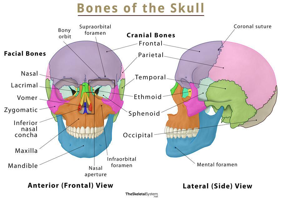
Anterior and Lateral View
Facial Skeleton
The facial skeleton, also known as the viscerocranium, comprises 14 facial bones (2 unpaired and 6 paired). Together, these bones structure our face, cheeks, nose, mouth, and jaws. Here are the bones in the facial skeleton:
- Maxillae/upper jaw bones (2)
- Lacrimal bone (2)
- Zygomatic bone/cheek bones (2)
- Palatine bone (2)
- Nasal bones (2)
- Inferior nasal concha (2)
- Vomer (1)
- Mandible (1)
Here, the hyoid bone, and the ear ossicles (middle ear bones) are also included in the facial bones. The three paired middle ear bones are malleus, incus, and stapes. Apart from forming the face, these bones are responsible for protecting and supporting the soft tissues in the area. The maxilla occupies most of the front part of the facial skeleton and forms the boundaries for the nasal aperture with the two nasal bones. The zygomatic, lacrimal, palatine, vomer, nasal bones, and the inferior nasal concha form the rest of the orbits.
Sutures of the Skull
Sutures are a unique type of fibrous joint that connect the skull bones. These joints allow for movement during infancy, so the brain and skull can grow. These joints fuse and become immovable around the age of 22-24.
As mentioned above, the three primary sutures in an adult skull are the ones between the cranium’s frontal, parietal, and occipital bones:
- Coronal suture – Between the frontal and the two parietal bones
- Sagittal suture – Between the two parietal bones
- Lambdoid suture – Between the occipital and the two parietal bones
In newborn babies, multiple soft spots or gaps exist between the skull bones due to the partially joined sutures. These gaps are known as fontanelles, with the frontal and occipital fontanelles being the most important ones. The soft spots gradually hardens and disappears as the sutures fuse in adults.
Important Foramina
The cranial base has multiple foramina and other openings to allow passage to the spinal cord, blood vessels, and cranial nerves. Here are the names of some of the most important foramina, along with the nerves they allow passage to:
- Foramen magnum in the occipital bone – the spinal cord
- Optic nerve canal in the sphenoid bone (the optic foramen is the opening to this canal) – Optic nerve and ophthalmic artery
- Foramen ovale in the sphenoid bone – Mandibular nerve (trigeminal nerve branch)
- Foramen rotundum in the sphenoid bone – Maxillary nerve (trigeminal nerve branch)
- Foramina spinosum in the sphenoid bone – Middle meningeal artery
- Cribriform plate in the ethmoid bone – Olfactory nerve
- Supraorbital and infraorbital foramina in the maxilla – Supraorbital and infraorbital nerves
- Mental foramina in the maxilla – Incisive and mental nerves and mental artery
- Supraorbital foramen in the frontal bone – Supraorbital nerve
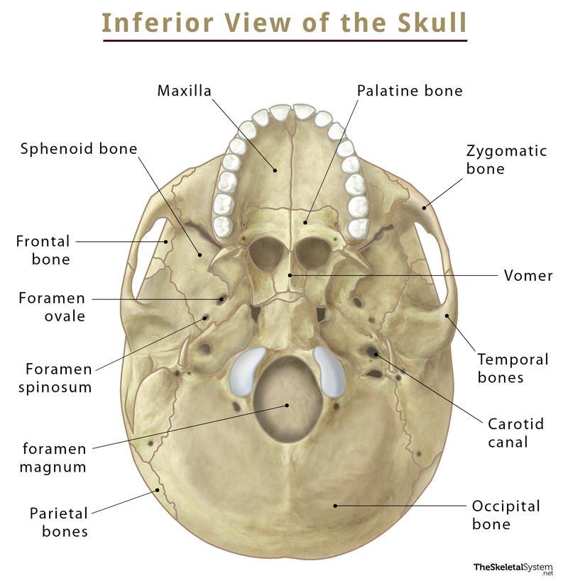
Inferior View of the Skull
Blood Supply
The skull primarily gets its blood supply from the common carotid artery, while the vertebral artery also contributes.
Muscles
The scalp and face muscles are innervated mainly by the facial, oculomotor, or trigeminal nerves. The hypoglossal nerve innervates the tongue. These muscles are responsible for everything from facial expressions, talking, and eating to lifting or lowering our eyebrows and moving our eyes to see.
FAQs
Q. Do skulls have bone marrow?
Ans. Yes, skull has bone marrow. Recent studies have found that the skull bone marrow is directly connected to the brain through special openings.
Q. What is the only movable bone in the skull?
Ans. The mandible or jawbone is the only movable bone in the skull.
Q. What bone protrudes at the base of the skull?
Ans. The occipital bone protrudes at the base of the skull.
Q. How thick and hard is a human skull
Ans. A female skull is slightly thicker than a male skull, with the former being 7.1 mm thick while the later has a thickness of 6.5 mm.When it comes to the hardness of human skulls, it can withstand a pressure of around 6.5 GPa. To compare this with some of the hardest objects, concrete can withstand 30 GPa, while steel up to 200 GPa.
References
- Bones of the Skull – Teachmeanatomy.info
- Skull – Kenhub.com
- Anatomy, Head and Neck, Skull – Ncbi.nlm.nih.gov
- Bones of the Skull – Joionline.net
- The Skull – Oregonstate.education
- Bones of the Skull – Libretexts.org