Pelvis
The pelvis is the lowermost part of the body trunk, located between the abdomen and the thighs. This basin-shaped bony structure protects a number of delicate organs, including the intestines and the reproductive systems.
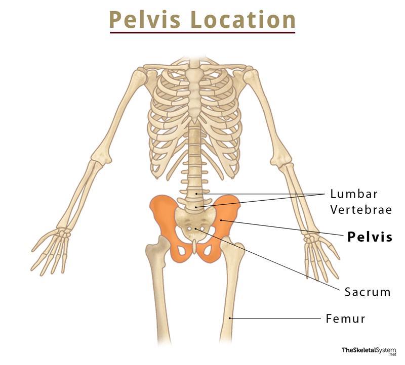
Pelvis Location
Names of the Pelvic Bones with Parts and Basic Anatomy
The pelvis can be divided into three parts – the bony pelvis, pelvic cavity, and the perineum.
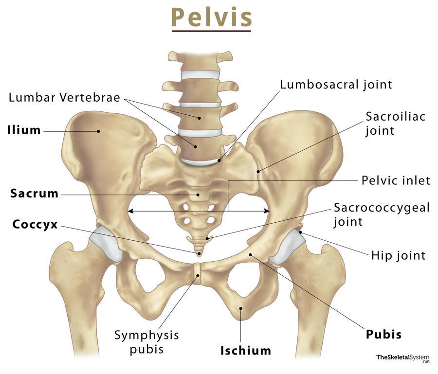
Pelvis Labeled Diagram
1. Bony Pelvis
As the name implies, this is the bony part of the pelvis, anatomically separated into two regions:
Pelvic Girdle
It is the ring-like part of the pelvis, formed by the following three fused bones:
These three bones fuse together to form the hip bone, which has a number of important landmarks, with the most prominent of them being the acetabulum. It is where the hip bone articulates with the femur to for the ball and socket hip joint.
The obturator foramen is an opening in the hip bone, encircled by the ischiam and pubis bones, located below and behind the acetabulum. This opening allows passage to a number of vital nerves and blood vessels that connect the hip region to the thigh and lower legs.
Pelvic Spine
This is the posterior part of the pelvis where the following bones of the pelvis are found:
The pelvic spine is located below the lumbar spine and is actually a part of the spinal column.So, even though these two bones play a vital part in structuring the pelvis, the sacrum and coccyx are counted in the spine bones, where they are even more important.
2. Pelvic Cavity
The large opening bounded by the three hip bones is called the pelvic cavity. It is continuous with the abdominal cavity, with the pelvic floor lying beneath it. Superiorly, the pelvic cavity opens into the abdomen through the superior pelvic aperture (pelvic inlet). The ischiopubic rami are located in the front, to the side of the inlet. The inferior opening of the cavity is known as the inferior pelvic aperture (pelvic outlet).
This opening is divided into the greater (false) and the lesser (true) pelvis. The greater pelvis is actually part of the intestines, as it is located above the pelvic inlet, holding the distal or last segment of the intestines.
The lesser pelvis on the other hand, lies between the pelvic inlet and outlet, containing the reproductive organs, internal genitalia, and the distal parts of the urinary tract.
3. The Perineum
It is the area below the pelvic floor, between the anus and the scrotum in males, and the anus and vagina in females.
Joints and Articulations
- Lumbosacral Joint: Between the L5 (fifth lumbar vertebra) and the sacrum
- Sacrococcygeal Joint: Amphiarthrodial joint between the fifth segment of the sacrum and the first segment of the coccyx
- Sacroiliac Joints: Between the sacrum (alae) and ilium on both sides of the body
- Symphysis Pubis: Between the left and right pubic bones through the articular surfaces of the two pubic bones.
- Acetabulofemoral Joint or Hip Joint: Between the acetabulum and the head of femur.
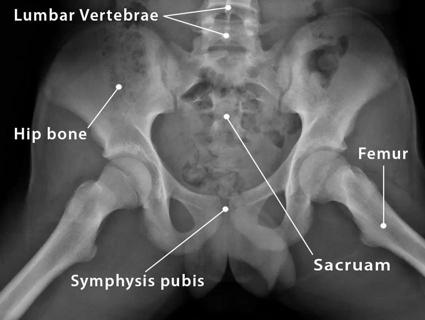
Pelvis X Ray
Functions
- Carries the upper body’s weight and distributes it from the axial skeleton (spine) to the appendicular skeleton of the thighs and legs.
- Allows us to move the lower part of the body, including the legs, so we can perform daily activities like walking, running, sitting, standing, etc.
- Protects various delicate organs like the pelvic colon, urinary bladder, rectum, and reproductive organs.
- Provides a point of attachment for several of the largest muscles in the body, including the levator ani, coccygeus, and puborectalis. These are necessary for locomotion, urination, defacation, and other such vital actions. The pubis is also the anchoring poit for a number of ligaments and small muscles from the pelvic floor and perineum.
- Provides a safe and comfortable space for the fetus to develop and grow during pregnancy. The female pelvis is also structured differently from males to to carry out labor and support childbirth.
- The pelvic floor and perineum regulate the urogenital and rectal openings.
Male Vs. Female Pelvis
The pelvic girdle may have different shapes in males and females. The superior pelvic aperture or pelvic inlet tends to be much wider in females, with shorter and relatively blunt ischial spines and wider ischiopubic rami. These structural differences work in favor of fetal growth and childbirth.
Males have a heart-shaped inlet, longer ischial spines projecting inward, and narrower ischiopubic rami.
Types of Pelvis
Though we have described the basic structural differences between male and female pelvis, more variations have been observed. So, human pelvises are classified into four categories.
- Gynecoid: The typical female pelvis.
- Android: The typical male pelvis.
- Anthropoid: Characterized by features of both the above types. Has increased risk of obstructed labor during pregnancy.
- Platypelloid: Also called contracted pelvis, it is similar to the gynecoid type, with a slightly curved sacrum.
Ossification and Development
The whole of the bony pelvis develops as cartilage, with the ilium, ischium, and pubis developing as separate bones. They remain separate at birth and through childhood.
Studies have shown that the female pelvis continues to grow wider, reaching its full width at the age of around 25-30 years. It starts narrowing again at around 40 years.
The male pelvis grows in the same manner till adulthood.
References
- Anatomy, Abdomen and Pelvis – Ncbi.nlm.nih.gov
- The Pelvis – Teachmeanatomy.info
- Bony pelvis – Kenhub.com
- Pelvic Anatomy – Hopkinsmedicine.org