Spine (Vertebral Column)
What is the Vertebral Column
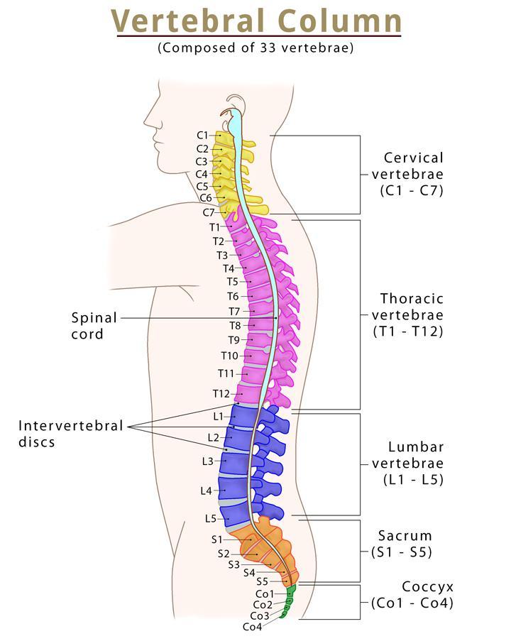
The vertebral column, commonly known as the spine, spinal column, or backbone, is a flexible hollow structure through which the spinal cord runs. It comprises 33 small bones called vertebrae, which remain separated by cartilaginous intervertebral discs. The vertebral column forms the axial skeleton, skull bones, ribs, and sternum.
Where is the Vertebral Column Located
The spine starts just below the occipital bone and extends up to the tip of the coccyx (tailbone).
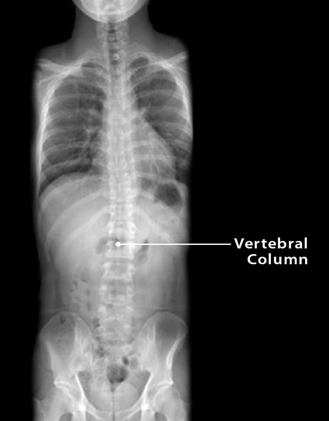
Vertebral Column X-ray
What are the Bones of the Spine
The 33 irregular vertebrae are categorized into the following 5 groups:
- Cervical vertebrae (7)
- Thoracic vertebrae (12)
- Lumbar vertebrae (5)
- Sacrum (5 fused)
- Coccyx (4 fused)
Spine (Vertebral Column)
Functions
- Protects the spinal cord from any mechanical injury by enclosing the cerebrospinal fluid (CSF), spinal cord, and nerve roots. Besides, it also protects various vital internal organs, such as the heart and lungs.
- Serves as the attachment point for several muscles, tendons, and ligaments, which are essential for body movement.
- Acts as the body’s central axis, bearing the whole body’s weight. It also supports the head, shoulder, and chest and balances the body by distributing the upper body’s weight to the lower extremities.
- Allows the spine to twist and bend through the joints between the vertebrae, the intervertebral discs. As a result, the spine can flex (bend forward), extend (bend backward), bend sideways, and rotate to some extent.
Parts and Anatomy
Though the anatomy of the 33 vertebrae may differ in some ways, most of them share a typical structure with the following parts:
1. Vertebral Body
It is the large, cylindrical, weight-bearing component that forms the anterior part of all vertebrae. Its superior and inferior regions are covered by hyaline cartilage. A fibrocartilaginous intervertebral disc separates the adjacent vertebral bodies from each other. As we move down the vertebral column, the size of the vertebral body increases to better support the body’s weight.
2. Vertebral Arch
It forms the lateral and posterior parts of each vertebra. The vertebral arch and vertebral body join to create an enclosed space called the vertebral foramen. These hole-like structures of all the adjacent vertebrae stack up on top of each other to form the continuous hollow space of the backbone, the vertebral canal. The spinal cord runs through this canal.
The vertebral arch features the following bony projections, where the muscles and ligaments get attached.
- Spinous processes
- Transverse processes
- Pedicles
- Lamina
- Articular processes
Parts of the Vertebral Column
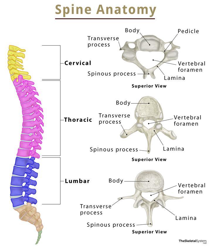
Spine Anatomy
1. Cervical Vertebrae
There are 7 cervical vertebrae named C1-C7, having thin intervertebral discs. The first two, C1 (atlas) and C2 (axis), have unique anatomy and help in the rotation of the head. The other five vertebrae, C3-C7, share similar anatomical features. They all have a bifid spinous process, transverse foramina, anterior and posterior tubercles, and a triangular vertebral foramen.
2. Thoracic Vertebrae
There are 12 medium-sized thoracic vertebrae (T1-T12) in the spine, with thicker intervertebral discs than cervical ones. The size of the individual vertebra increases gradually down the spine. Their job is to articulate with the bony ribs, forming the rib cage or thoracic cage.
3. Lumbar Vertebrae
The spine has 5 lumbar vertebrae(L1-L5) supporting the body weight. They have large, kidney-shaped vertebral bodies, triangular vertebral foramen, and short spinous processes. Among all the vertebrae, the L5 lumbar vertebra is the largest.
4. Sacrum
It looks like a single bony component composed of 5 fused vertebrae (S1-S5). It looks like an inverted triangle, with the apex pointing inferiorly.
5. Coccyx
It is another component of the vertebral column, formed by 4 fused vertebrae (Co1-Co4). It articulates with the apex of the sacrum and is devoid of the vertebral canal.
Spinal Curves
The spine in adults has multiple natural curves, with the thoracic and sacrococcygeal curves being the primary ones. There are two other minor curvatures, the cervical, and lumbar curves, at the respective regions of the spine.
These curves provide extra strength to the spine to support and distribute the body’s weight, absorb shock, and allow the spine to bend and move in different directions.
Articulations
- Intervertebral symphyses: All the vertebral bodies, except C1-C2 and S2-S3, are joined via fibrocartilaginous joints by intervertebral fibrocartilage in the form of intervertebral discs.
- Zygapophyseal joints: Synovial joints formed between the adjacent vertebral arches’ superior and inferior articular facets.
- Atlanto-occipital joint: Another synovial joint found between the atlas (C1) and the occipital bone.
Ossification
The ossification of every vertebra begins around the 8th week of the gestation period. Vertebrae arise from three primary ossification centers: one for the vertebral body and two for the pedicles.
Muscle and Ligament Attachments
Muscle attachments
- Lumbar muscles
- Thoracic muscles
- Back muscles
Ligament attachments
- Ligamenta flava
- Interspinous ligaments
- Nuchal ligament
- Supraspinous ligament
References
- The Vertebral Column — Teachmeanatomy.info
- Anatomy, Back, Vertebral Column — Ncbi.nlm.nih.gov
- Vertebral column (spine) — Kenhub.com
- Vertebral Column — Sciencedirect.com
- The Vertebral Column — Courses.lumenlearning.com