Ilium
What is the Ilium Bone
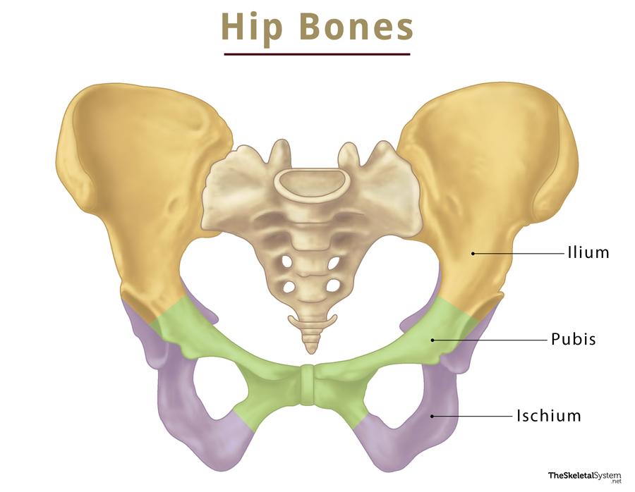
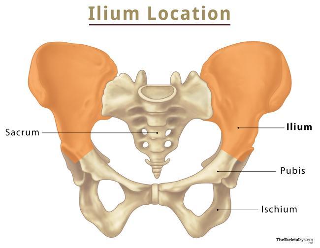
Ilium (plural: ilia), also known as the iliac bone, is one of the three bones that fuse to form the hip bone. The other two are the ischium and pubis. This largest and uppermost bone of the hip is an essential part of the pelvic girdle.
Where is the Iliac Bone Located
It is located on the uppermost portion of the hip bone. If you firmly press on your waists, you can feel the ilium, particularly the ilium crest.
Quick Facts
Functions
- Forms a part of the pelvis, thus protecting the reproductive organs, urinary bladder, and lower part of the digestive tract that lie within it.
- Bear body weight while resting or moving.
Parts and Anatomy of Ilium
This blade-shaped bone consists of two main parts: the body and ala (wing).
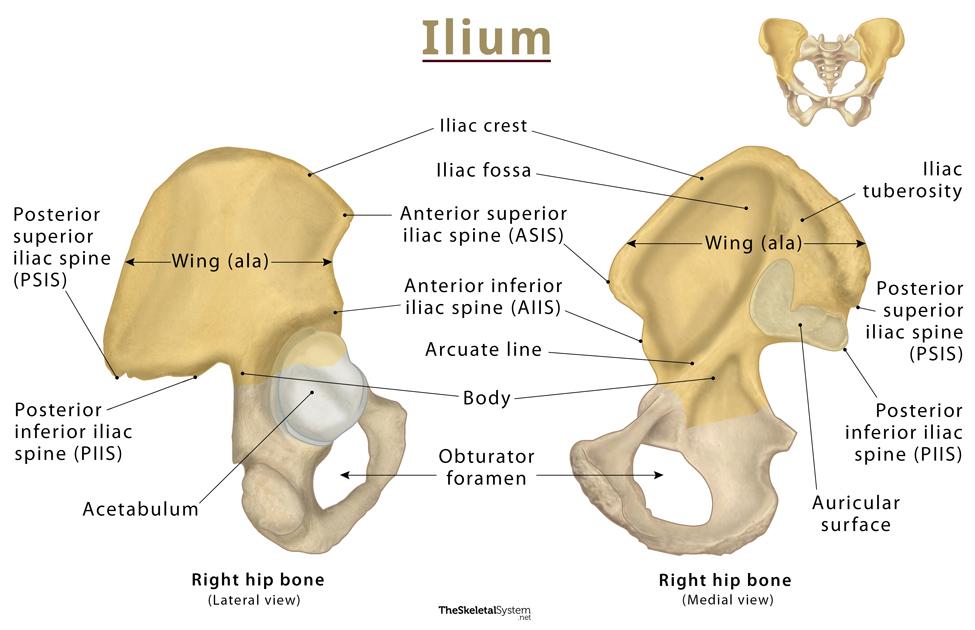
1. Body: The body is the smaller, inferior part of the bone that forms the upper boundary of the acetabulum. The internal surface of the body is part of the wall of the lesser pelvis, from where some fibers of the obturator internus muscle originate. The external surface is partly articular, forming the lunate surface of the acetabulum. The non-articular portion of the external surface contributes to the acetabular fossa. The lower portion of the body is continuous with the pelvic surfaces of the ischium and pubis. A faint line indicates the site of this union of these three bones.
2. Ala (Wing): It is the large, expanded portion of the bone which laterally bounds the greater pelvis. The superior border of the wing is thickened, creating the iliac crest. It extends from the anterior superior iliac spine (ASIS) to the posterior superior iliac spine (PSIS). On the posterior side, there is an indentation known as the greater sciatic notch. The inner surface of the wing has a concave shape, giving rise to the iliac fossa, the site of origin for the iliacus muscle. On the other hand, the external surface of the wing has a convex shape and provides attachments to the gluteal muscles.
Landmarks
The ilium has four major protruding areas or iliac spines, which are important bony landmarks.
- Anterior Superior Iliac Spine (ASIS): It is located at the anterior end of the iliac crest and serves as a point of attachment for the inguinal ligament.
- Anterior Inferior Iliac Spine (AIIS): It is located anterior to the supra-acetabular groove and acetabular margin. AIIS is separated from the ASIS by a short, vertical slope. It provides attachment points for the rectus femoris and proximal part of the iliofemoral ligament.
- Posterior Superior Iliac Spine (PSIS): This spine is located at the posterior end of the iliac crest and is laterally related to the iliac tuberosity and sacropelvic surface. It is commonly represented by a dimple above the medial gluteal region
- Posterior Inferior Iliac Spine (PIIS): It is located inferior to the PSIS.
Borders
The ilium features four distinctive borders; superior (iliac crest), anterior, posterior and medial.
1. Superior Border (Iliac crest)
The superior border of the ilium is known as the iliac crest. It is a rough, crescentic surface that starts at the PSIS posteriorly and arches forward, ending at the ASIS anteriorly. The iliac crest features an inner and an outer lip, with the zone between the two marked as the intermediate zone. The superior border is a site of attachment for several muscles and fascia of the abdominal wall, back, and lower limb.
2. Anterior Border
The anterior border of the ilium stretches from ASIS to the acetabulum. It bears AIIS just superior to its acetabular end. The part of the border that runs between these spines is concave anteriorly.
3. Posterior Border
The posterior border of the ilium begins at the posterior superior iliac spine and extends to the posterior border of the ischium. It features a posterior inferior iliac spine and contributes to the superior part of the greater sciatic notch. The course of this border is irregular; the part between the spines is concave posteriorly, while the part from the inferior spine to the ischial border first runs horizontally and then posteroinferiorly to comprise the superior border of the greater sciatic notch. This notch is completed inferiorly by the posterior ischial border and ischial spine. The sacrospinous and sacrotuberous ligaments enclose the notch superiorly and posteroinferiorly, converting it into the greater sciatic foramen.
4. Medial border
It features a smooth, rounded line, called the arcuate line, running anteroinferior from the auricular surface to the acetabulum. This is the point where the body and ala join.
Surfaces
The four borders of the ilium surround the three bony surfaces; gluteal, sacropelvic, and iliac.
1. Gluteal Surface
The posterolateral surface is the gluteal surface that allows several gluteal and thigh muscles to attach to the bone. The iliac crest is located above this surface. There are three curved ridges, the anterior, posterior, and inferior gluteal lines. The supraacetabular groove is another important landmark located between the acetabular margin and the inferior gluteal line. It is the point of attachment for the reflected head of the rectus femoris muscle.
2. Sacropelvic Surface
The sacropelvic surface, is composed of the iliac tuberosity, auricular, and pelvic surfaces. It is the medial aspect of the bone located at the back of the iliac fossa. This is where the ilium articulates with the sacrum at an ear-shaped surface to form the sacroiliac joint. The iliac tuberosity is a rough, elevated area at the back of this surface, where the ligaments of the sacroiliac joint attach. Another surface, called the pelvic surface, is there below and at the front of the articular surface — it contributes to the formation of the lateral wall of the lesser pelvis.
3. Iliac (internal) Surface
The large concave surface located medially and in front of the wing of the ilium is called the iliac fossa. Its main purpose is to form the smooth walls at the back and sides of the greater pelvis. It is separated from the sacropelvic surface by the medial border.
Articulations
Sacroiliac joint: It is a synovial joint formed between the ilium and the sacrum
Muscle and Ligament Attachments
Several important muscles and ligaments originate or insert onto the ilium.
Muscle Attachments
Muscles that originate from ilium:
- Sartorius muscle: At the anterior superior iliac spine.
- Rectus femoris muscle: From the anterior inferior iliac spine, the reflected head of this muscle originates from the supra-acetabular region of the ilium.
- Gluteus maximus, medius and minimus muscles: From the gluteal surface.
- Iliacus muscle: From upper two-thirds of the iliac fossa.
- Tensor fascia lata: From the anterior and dorsal aspect of the iliac crest.
Muscles that insert onto the ilium:
- Quadratus lumborum muscle
- External oblique
- Internal oblique
- Transversus abdominis muscles
- Latissimus dorsi
All of these muscles insert at the iliac crest.
Ligament Attachments
- Inguinal ligament: At the anterior superior iliac spine.
- Iliofemoral ligament: At the anterior inferior iliac spine.
- Sacrotuberous ligament: At the posterior inferior iliac spine.
- Posterior sacroiliac ligament: At the iliac tuberosity.
- Interosseous sacroiliac ligament and ventral sacroiliac ligament: At the auricular surface of iliac tuberosity.
- Iliolumbar ligament: At the anterior side of iliac tuberosity.
References
- Ilium – Radiopaedia.org
- The Hip Bone – Teachmeanatomy.info
- Ilium – Med.libretexts.org
- Hip bone – Kenhub.com
- Ilium (Bone) – Sciencedirect.com