Coccyx (Tailbone)
Published on December 31st 2021 by staff
What is the Coccyx
The coccyx is the smallest and the most inferior bone in the spinal column. It is the terminal part of the vertebral column that makes up the very bottom portion of the spine below the sacrum. It is usually composed of four vertebrae (Co1- Co4), which fuse to produce a triangular shape, resembling a shortened tail. As it is the remnant of the vestigial tail, it is commonly known as the tailbone. The coccyx is one of the bones that bear the body’s weight while sitting.
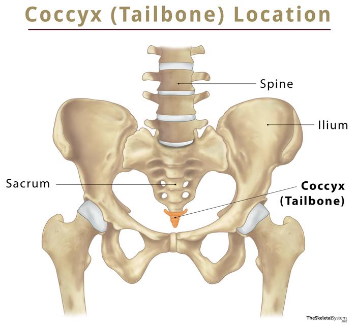
Where is the Coccyx or Tailbone Located
The coccyx is located in the most distal portion of the spine, just below the sacrum. It is the lowermost part of the pelvic girdle or pelvis.
Quick Facts
| Type | Irregular bone |
| Length | Approximately 1 inch |
| How many are there in the human body | 1 |
| Articulates with | Sacrum |
Functions
Bears body weight while sitting: When a person is seated, the body weight gets distributed between the bottom portions of the two hip bones or ischium and the tailbone, thus providing balance and stability.
Anatomy
Coccyx looks like an inverted triangle, having a wide base at the top, and a pointy apex at the bottom. It also bears several other important bony landmarks.
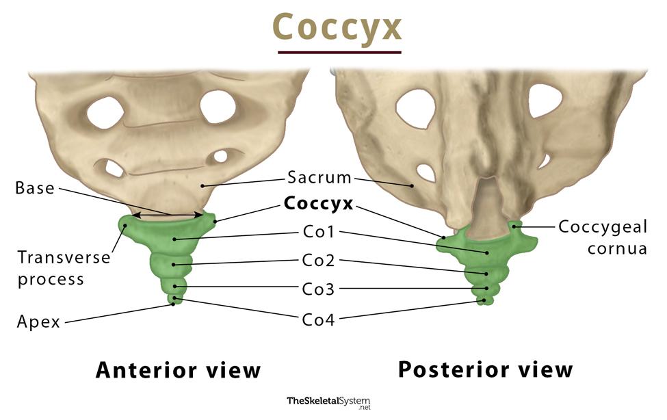
Bony Landmarks
Along with the base and apex, it also has four surfaces, anterior surface, posterior surface, and two lateral surfaces.
Anterior Surface
The anterior surface of the coccyx is slightly concave and marked with three transverse grooves, indicating the junctions of the four vertebrae. The anterior sacrococcygeal ligament gets attached here.
Posterior Surface
Similar to the anterior surface, the posterior surface is also marked by three transverse grooves. Unlike anterior, the posterior surface is convex, presenting a linear row of tubercles on either side. Tubercles are the rudimentary articular processes of the coccygeal vertebrae. Out of these, the superior pairs are the largest and are called the coccygeal cornua. They project upwards to articulate with the sacral cornua. The coccygeal and sacral cornua combine to form a foramen for the transmission of the posterior division of the fifth sacral nerve.
Lateral Surface
The coccyx has two lateral surfaces on either side, bounded by lateral borders. The lateral borders are thin, and have a series of small eminences, representing the transverse processes of the coccygeal vertebrae. Out of these, the first one is the largest. It is flattened and often ascends to join the lower part of the thin lateral edge of the sacrum. This articulation forms a foramen through which the anterior division of the fifth sacral nerve passes. The rest of the eminences or transverse processes gradually decrease in size and diminish.
The lateral borders of the coccyx serve as an attachment site for the sacrotuberous, coccygeus, and sacrospinous ligaments, and the fibers of the gluteus maximus muscle.
Base
The top, wide portion of the coccyx is referred to as its base. It contains a proximal, oval facet for articulation with the sacrum.
Apex
The rounded terminal end of the bone is called its apex. The tendon of the external anal sphincter gets attached here.
Articulations
1. Sacrococcygeal Joint: The coccyx articulates with the sacrum, forming a fibrocartilaginous joint. The joint can move to a limited extent, i.e., minor flexion and extension, which occurs passively during defecation and labor.
Ossification
At birth, the coccyx arises as a cartilaginous mass attached to the end of the sacrum, which gradually ossifies as individual bones. The ossification happens from top to bottom. The process gets completed by the end of puberty. Initially, the four vertebrae remain separate, but they fuse throughout adulthood to form one continuous bone.
Anatomical Variations of the Coccyx
Coccyx shows considerable anatomical variation between individuals.
- One typical variant is the failure of the first coccygeal vertebra (Co1) to fuse with other vertebrae. Thus it remains separate throughout adult life. In some cases, the four vertebrae may only fuse partially in some individuals, resulting in the coccyx consisting of two separate bones.
- In some individuals, instead of 4, the coccyx can also be composed of one less or one more coccygeal vertebra, i.e., 3 or 5 vertebrae.
- In more than half of adults (about 57%), the joint between the sacrum and the coccyx remains fused.
Coccyx in Females vs. in Males
Coccyx also features variation in its shape and curvature, which vary between individuals and is noticeably different between sexes.
The anterior surface of the coccyx is concave, giving a curved appearance to it. This concave surface is continuous with the curvature of the sacrum. In females, this concavity or curvature is reduced, while in males, it is greater. So, in females, the coccyx does not point as anteriorly as it does in males. Also, there is a noticeable difference between the shapes of the coccyx in males and females. The female coccyx is narrower and less triangular.
Muscle and Ligament Attachments
Muscles
Coccyx serves as an attachment point for various muscles.
Muscles attached to the anterior side of the coccyx:
- Levator ani muscle
- Coccygeus
- Iliococcygeus
- Pubococcygeus
- Anococcygeal raphe
Muscles attached to the posterior side of the coccyx:
- Gluteus maximus
Ligaments
The sacrococcygeal joint is supported by five ligaments:
- Anterior sacrococcygeal ligament
- Deep posterior sacrococcygeal ligament
- Superficial posterior sacrococcygeal ligament
- Lateral sacrococcygeal ligaments
- Interarticular ligaments
FAQs
Ans. The coccyx is vestigial, as, through evolution, it has lost its function as a tail.
Ans. The coccyx is a part of the axial skeleton.
References
- The Coccyx – Teachmeanatomy.info
- Anatomy, Back, Coccygeal Vertebrae – Ncbi.nlm.nih.gov
- Coccyx – Radiopaedia.org
- Coccyx – Innerbody.com