Rib Cage
What is the Rib Cage
The rib cage, also known as the thoracic cage, is the bony structure that shapes and protects the thoracic cavity and the organs in it. It has a conical shape and looks somewhat like a birdcage (hence the name). This part of the axial skeleton is located in the chest, above the abdominal cavity.
Functions
- The rib cage protects the two major organs in the thoracic cavity, the heart and the lungs.
- Supports the upper body, keeping it stable.
- It provides a point of attachment for the clavicles, thus establishing the connection between the arm and the axial skeleton.
- Being such a unique bony structure, it provides points of attachment for several pectoral girdle muscles, and core stability muscles.
- Stabilizes the thoracic cavity through its measured outward and inward movements during inhalation and exhalation.
Names and Anatomy of Bones in the Rib Cage
The human rib cage comprises 37 bones:
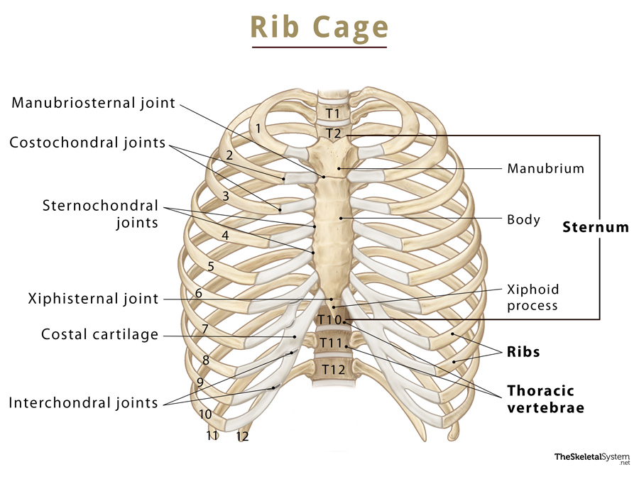
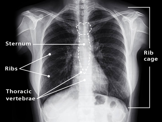
- Sternum (1, unpaired): It is also known as the breastbone and can be felt at the center of the chest. This bone articulates with the first 7 ribs.
- Thoracic vertebrae (12, unpaired): These are the 12 bones in the thoracic spine (T1-T12). Each vertebra articulates with its corresponding ribs to shape the rib cage. The thoracic spine’s primary function is to support and stabilize the rib cage.
- Ribs (12, paired): These long, curved, flat bones enclose the front of the rib cage. Out of the 12 paired bones, the first 7 pairs articulate with the sternum through their respective costal cartilages.
The gap between two ribs is called the intercostal space. Since there are 12 pairs of ribs, there are 11 intercostal spaces on each side of the rib cage, with each space named based on its location. For example, the 1st intercostal space would be located between the 1st and 2nd ribs. These spaces hold the intercostal muscles and help expand the rib cage during inhalation.
The opening at the bottom of the rib or thoracic cage is referred to as the inferior thoracic aperture or thoracic inlet. In contrast, the top opening is called the superior thoracic aperture or thoracic outlet. These two openings allow the organs inside the rib cage to connect with the systems and structures outside.
Articulations
- Xiphisternal joint – Between the sternum’s body and xiphoid process
- Manubriosternal joints – Between the sternum’s body and the manubrium
- Sternoclavicular joints – Between the manubrium (top of the sternum) and the clavicles
- Sternochondral joints – Between the sternum and the costal cartilage
- Costochondral joints – Between the ribs and their costal cartilage
- Costovertebral joints – Between the ribs and the thoracic vertebral bodies.
- Intervertebral joints – Between the 12 thoracic vertebrae
- Interchondral joints – Between the costal cartilages.
Muscle Attachments
The following muscles are located in the rib cage and help with its functioning:
- Intercostals (external, internal, and innermost layers)
- Subcostals
- Transversus thoracis
References
- Thoracic cage: KenHub.com
- The Thoracic Cage: Bones, Structure & Classifications: Study.com
- Rib Cage: What To Know: WebMD.com
- Rib cage: Anatomy.app
- The Thoracic Cage: OregonState.education