Thoracic Vertebrae (Thoracic Spine)
Published on May 24th 2022 by staff
What is Thoracic Spine
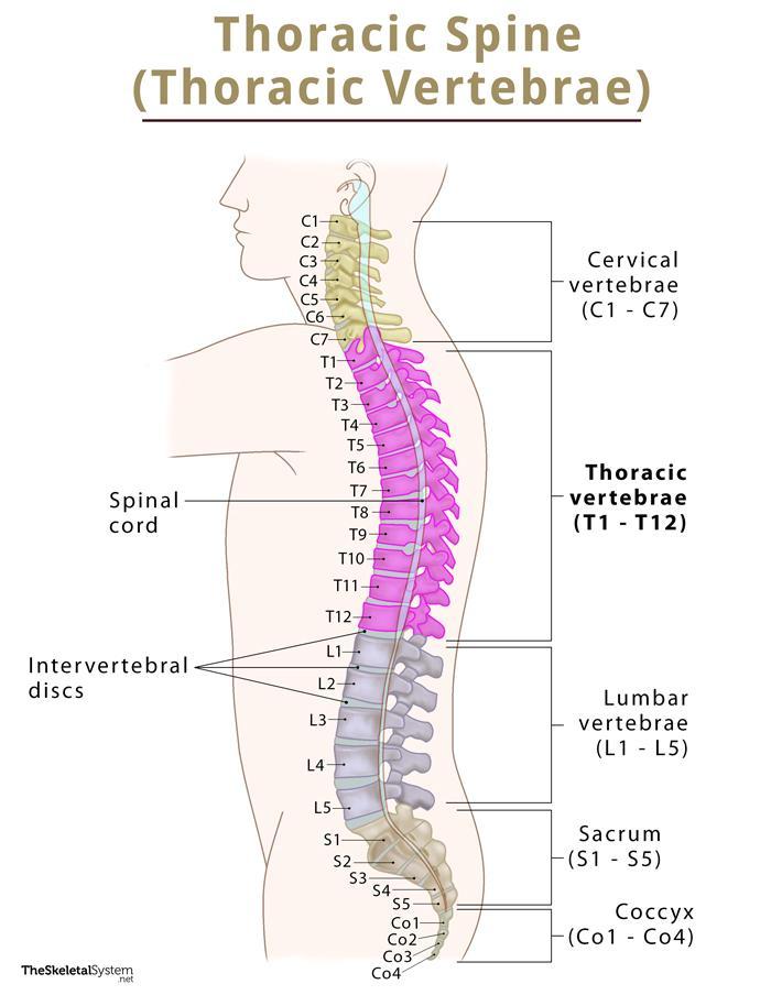
The thoracic spine is the second and longest part of the spinal column, consisting of 12 lumbar vertebrae, T1-T12. These 12 bones are separated from each other by intervertebral discs. Their primary role is to form the thoracic cage that protects the heart, lungs, and esophagus.
Where are the Thoracic Vertebrae Located
The thoracic vertebrae are stacked between the cervical and lumbar vertebral segments. They are found in the thoracic region, almost in the middle of the back.
Functions
- Protecting the spinal cord and providing its passage through the tunnel at the center of the vertebral column as the vertebrae stack one after another.
- Articulating with the ribs. All the thoracic vertebrae, except the two at the bottom (T11 and T12), form these articulations.
- Forming the rib cage and keeping it stable, protecting all the delicate internal organs, mainly the heart, and lungs.
- Supporting the body and allowing for movement; however, this part has the least range of movement in the entire spinal column.
Anatomy of Thoracic Vertebrae
The thoracic spine consists of typical vertebrae with all the individual bones having similar structure.
Vertebral body
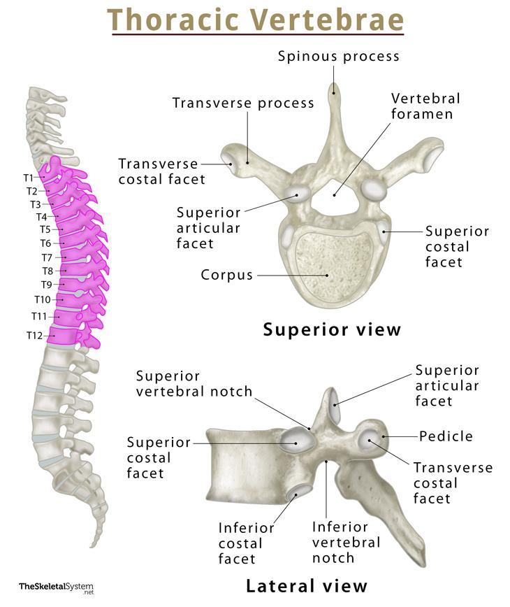
Thoracic vertebrae have medium-sized and heart-shaped vertebral bodies, also known as corpus. Their primary role is to support body weight. As it approaches the lumbar vertebrae, the size of the vertebral bodies increases. On both sides of the vertebral body lie two concave cartilage-lined depressions, named superior and inferior costal facets, where the ribs get attached. Among the two facets, the superior one articulates with the head of the adjacent rib, and the inferior articulates with the head of the rib below.
Pedicles
Pedicles are cylindrical bony protrusions projecting from the posterolateral surfaces of the vertebral bodies. Their superior and inferior surfaces are marked with several notches that combine to form the intervertebral foramina. The thoracic spinal nerves pass through this foramen. On the posterior side, the pedicles articulate with the laminae on both sides, forming the neural arch. This arch fuses with the posterior surface of the vertebral body, creating the vertebral foramen. The foramen of the adjacent thoracic vertebrae line up to form the vertebral canal, through which the spinal cord passes.
Laminae
Laminae arise from the posterior side of the pedicles and extend towards the posterior midline, forming the spinous process.
Transverse Processes
These are long and thin wing-like structures projecting laterally from the junction between the pedicle from both sides of the vertebrae. The tubercle of the rib, along with several essential muscles and ligaments, attach here.
Articular Processes
Every thoracic vertebra features superior and inferior articular processes on both sides. Each of these processes has its corresponding articular facets, superior articular facet, and inferior articular facet. The former projects posteriorly and laterally, while the latter directs forward and medially.
Articulations
- Intervertebral symphyses: The facet joints where the individual vertebra articulate with each other through intervertebral discs.
- Costovertebral Joints: Synovial joints formed between the proximal head of the rib and its corresponding vertebrae.
- Costotransverse Joints: Another group of synovial joints found between the rib’s tubercle and the corresponding vertebra’s transverse process.
Muscle and Ligament Attachments
Several muscles and ligaments attach to the thoracic vertebrae.
Muscles attached
- Erector spinae
- Interspinales
- Intertransversarii
- Latissimus dorsi
- Multifidus
- Rhomboid major
- Rhomboid minor
- Rotatores
- Semispinalis
- Serratus posterior (superior and inferior)
- Splenius capitis
- Splenius cervicis
- Trapezius
Ligaments attached
- Anterior longitudinal ligament
- Supraspinous ligament
- Posterior longitudinal ligament
- Ligamentum flavum
References
- The Thoracic Spine — Teachmeanatomy.info
- Anatomy, Back, Thoracic Vertebrae — Ncbi.nlm.nih.gov
- Thoracic vertebrae — Kenhub.com
- Typical thoracic vertebrae — Radiopaedia.org
- Thoracic Vertebrae — Innerbody.com