Ribs
Table of Contents:
Published on November 18th 2022 by staff
What Are Rib Bones
The ribs are 12 pairs of curved, flat bones that form the thoracic cage or rib cage, the bony structure that shapes the thoracic cavity and protects various organs. Despite being relatively thin and light, these bones are highly resilient.
Where Are the Ribs Located
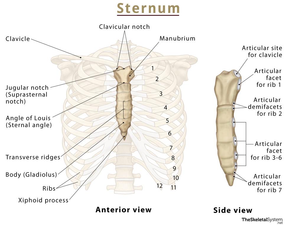
Ribs are located in the chest. They attach to the sternum or breastbone at the front and the thoracic spine in the back. The ribs can be felt and seen both at the front and back of the upper body. They are part of the axial skeleton.
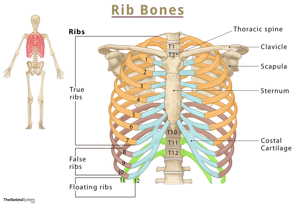
The ribs are numbered from 1 to 12 from top to bottom to match the numbering of their corresponding thoracic vertebra.
Quick Facts
| Type | Flat bone |
| How many are there in the human body | 24 (12 pairs) |
| Articulates with | Thoracic vertebrae |
Functions
As the ribs form the ribcage, their primary function is to enable it to work in order.
- The ribs surround the chest cavity and protect the heart and lungs, the two vital organs here.
- They facilitate respiration by moving both to front and back, and up and down to accommodate the lungs as they expand and contract during inhalation and exhalation. The diaphragm and intercostal muscles control these movements.
- They also provide points of origin or attachment for several essential muscles in the chest and thoracic wall.
- Rib bones participate in erythropoiesis (the process of producing red blood cells). They are one of the sites where erythropoiesis occurs throughout your adult life.
Anatomy
Types of Ribs
The 24 ribs are classified in two different ways. There are three types of ribs based on their attachment to the sternum:
- True ribs are the first 7 pairs that attach to the thoracic vertebra in the spine and then directly articulate with the sternum through their costal cartilage.
- False ribs are the next 3 pairs (8th, 9th, and 10th ribs). Their costal cartilages join with that of the 7th rib to indirectly connect to the sternum.
- Floating ribs are the last 2 pairs (11th and 12th ribs) with no articulation with the sternum.
The second mode of classifying ribs is based on their structure and anatomy variations. Based on this, they can be of two types — typical and atypical.
Typical Ribs
The 3rd to 9th pairs of ribs are the typical ribs as they all have a typical structure with the following features and landmarks:
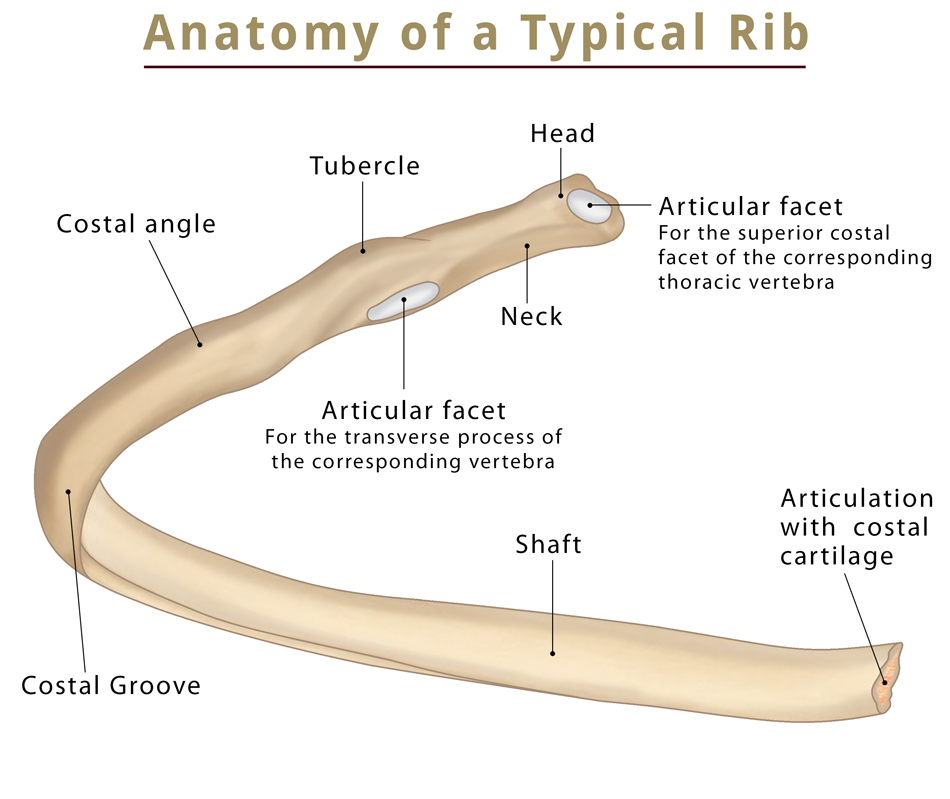
Head: It is the wedge-shaped (medioposterior) end of a rib bone where two articular surfaces are divided by a bony ridge. The larger one of the two surfaces articulates with the superior costal facet of the corresponding thoracic vertebra, while the smaller one with the inferior costal facet of the vertebra above.
For example, the larger articular surface of the third rib attaches with the T3 (second thoracic vertebra) and the smaller surface with the T2.
Neck: This part connects the head to the rib shaft. There are no bony markings here.
Tubercle: The rough bony protrusion where the neck and shaft meet. The only articular surface in this region is for the transverse process of the corresponding vertebra.
Shaft: The long, thin curved part of the rib following the tubercle. The curve towards the front of the body is most pronounced at the costal angle. This angle marks the point of attachment for several deep back muscles.
At the shaft’s end is one cup-shaped surface where the rib attaches with costal cartilage.
Costal Groove: The groove or depression along the inferior border. It allows passage to the neurovascular bundle that includes the intercostal blood vessels and nerves.
Atypical Ribs
These are the ribs that have characteristic features not present in other ribs.
1st Rib: This short and thick rib has only one articular surface for the T1 vertebra. It has a head, neck, and body but does not curve as sharply as the typical ribs and has no costal groove. The superior surface is marked with two grooves, with a small ridge in between allowing passage to the subclavian blood vessels. It is also the first point of origin of the serratus anterior muscle.
2nd Rib: Though smaller than a typical rib, it is longer and thinner than the 1st rib, with two articular facets on the head for T1 and T2. Its main distinguishing feature is the rough tuberosity on its upper surface which acts as one of the points of origin of the serratus anterior muscle.
10th Rib: It has only a single articular surface for its corresponding vertebra (T10).
11th and 12th Ribs: Like the 10th rib, they also have a single facet for their corresponding vertebra. Additionally, these two ribs are very short, with no neck or tubercle.
Articulations
- Costovertebral joints: Connects the head of the rib to the corresponding thoracic vertebrae, and the one above it
- Costochondral joint: Connects the costal groove of a rib to its corresponding costal cartilage
- Costotransverse joint: Connects the tubercle to the transverse process of the corresponding vertebra
- Sternocostal joint: Connects the true ribs to the sternum or breastbone
- Costoclavicular joint: An anatomical variant where the 1st rib connects with the clavicle
Muscle Attachments
Several vital muscles attach to the ribs, controlling or affecting their movement. Here are the names of some of the most important muscles around the ribs:
- Intercostals (external, internal, innermost)
- Subcostales
- Transversus thoracis
- Serratus posterior
- Serratus anterior
- Levatores costarum
- Pectoralis major
- Pectoralis minor
- Latissimus dorsi
- Rectus abdominis
FAQs
Ans. There is no difference in the number of ribs in men and women. All humans have 12 pairs of ribs.
Ans. The primary difference between true and false ribs is that the former attaches to the sternum directly, while the latter attaches to the sternum indirectly (through the 7th costal cartilage).
Ans. Studies have shown that ribs and costal cartilage can regenerate as long as the connective tissues surrounding them are intact. The ribs are surrounded by the vascular connective tissue perichondrium, while the periosteum surrounds the cartilages.
Ans. A cervical rib is a rare anatomical variation present at birth in 0.5-1% of people with an extra rib present above the 1st rib. It might grow on one or both sides, either as a bony outgrowth or as a fully formed rib. Cervical ribs are more common among women than men.
It often does not cause any problems in the individual. Still, in some cases, the bony growth might press on the nerves and blood vessels in the area, causing neck pain and weakness, numbness, or dislocation of the arm (thoracic outlet syndrome).
References
- The Ribs: TeachMeAnatomy.info
- Ribs: KenHub.com
- Anatomy, Thorax, Ribs: NCBI.nlm.nih.gov
- Typical Ribs: RadioPaedia.org
- Structure of the Ribcage and Ribs: GetBodySmart.com
- 3D Skeletal System: Bones of the Thoracic Cage: VisibleBody.com