Hip Bone (Coxal Bone)
What is the Hip Bone
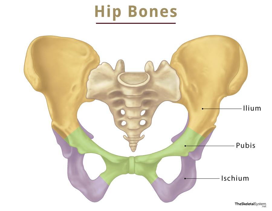
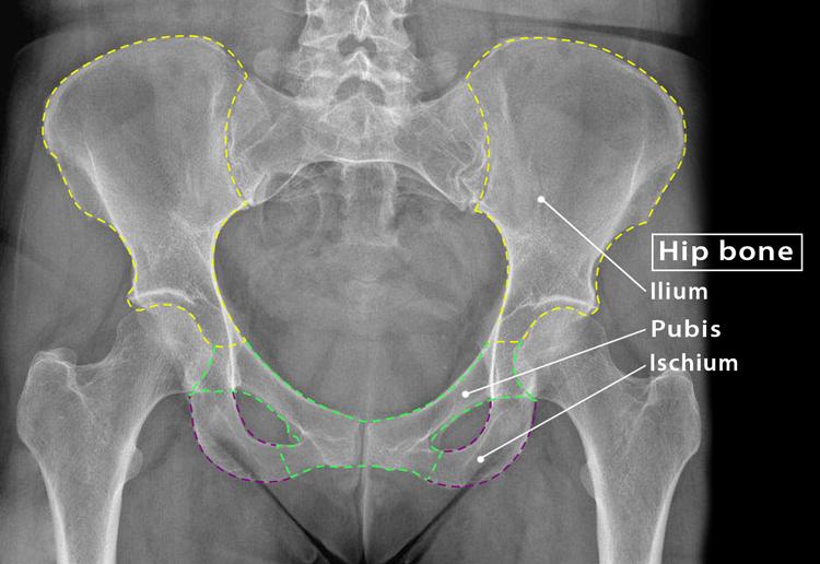
Hip bone, also known as the coxal bone, innominate bone, or pelvic bone, is an irregular bone found on both sides of the body. These left and right hip bones join to form the pelvic girdle, where the delicate organs of the lower abdomen are found. Though it looks like a single bone, it comprises three fused bones: ilium, ischium, and pubis.
Where is the Hip Bone Located
The hip bone is located on each side of the pelvis. Anatomically, it is positioned just below the backbone or spinal column. You can easily feel these bones by touching the sides of your hip region.
Quick Facts
| Type | Irregular bone |
| How many are there in the human body | 2 (1 on each side) |
| Articulates with | Sacrum and femur |
Functions
- Form the pelvis, along with the sacrum and coccyx, where the delicate organs like kidneys, urinary bladder, a part of the lower intestine, and reproductive organs lie.
- Connect the lower limbs with the axial skeleton.
- Support the body weight while sitting or moving.
Parts and Anatomy
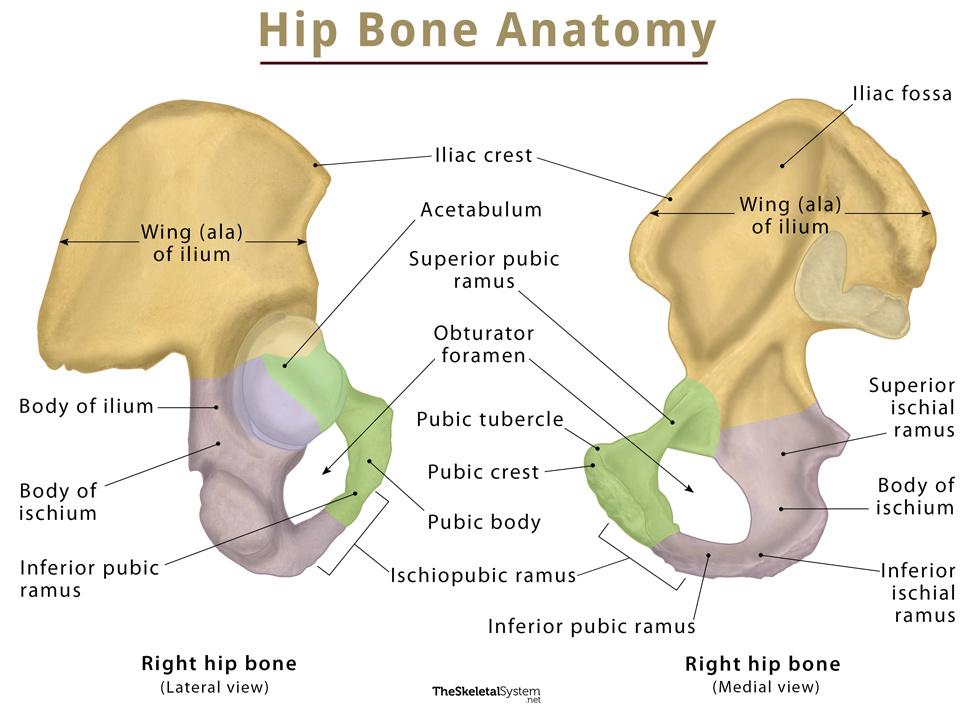
Three separate bones, ilium, ischium, and pubis, fuse to form the pelvic bone. These three bones give rise to a cup-shaped socket called the acetabulum. Hip bones also feature another distinguishing landmark, obturator foramen.
Important Landmarks
Acetabulum
As mentioned, the three bones of the hip unite to form this prominent bony landmark, present on the lateral sides. It is a cup-shaped socket-like structure featuring a moon-shaped articular surface, called lunate surface for the head of the femur. So, here, the head of the femur fits into the acetabulum, forming the hip joint.
Obturator foramen
At the anteroinferior side of the acetabulum, there is a large obturator foramen bounded by the pubis and ischium. The major portion of this foramen remains covered by a flat connective tissue membrane called the obturator membrane.
Bones That Form the Hip Bone
As we know, the three bones, ilium, ischium, and pubis, form the hip bone. The ilium is the largest and most superior part of the bone, the ischium forms the posteroinferior side, and the pubis or pubic bone forms the anterior aspect. Below is a quick overview of these bones:
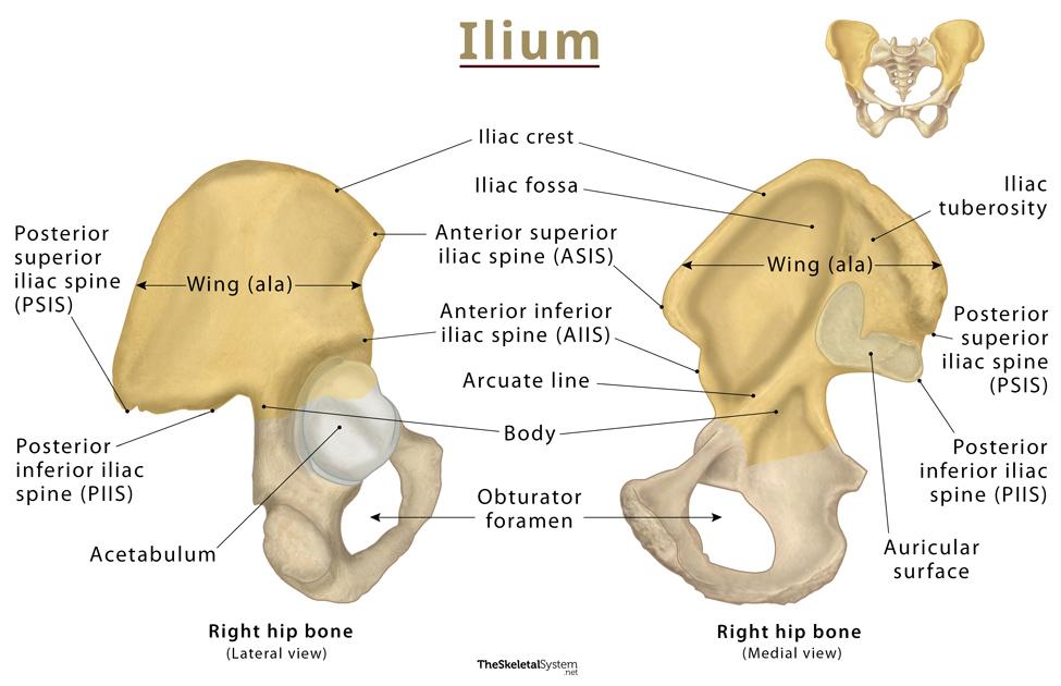
Ilium
It is a blade-shaped bone comprising two parts: the body, and ala or wing. The body is the smaller, inferior part, while the ala is the superior expanded part of the bone. The body of the bone contributes to the formation of the acetabulum. The superior border of the wing is thickened, giving rise to the iliac crest.
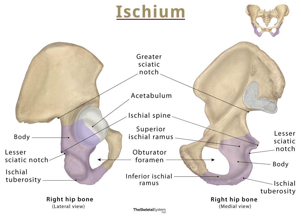
Ischium
It is the posterior and inferior part of the bone, which is divided into two main parts: the body, and two rami (superior ramus and inferior ramus). The body is the largest portion of the bone, projecting upwards to join the ilium and the superior ramus of the pubis.
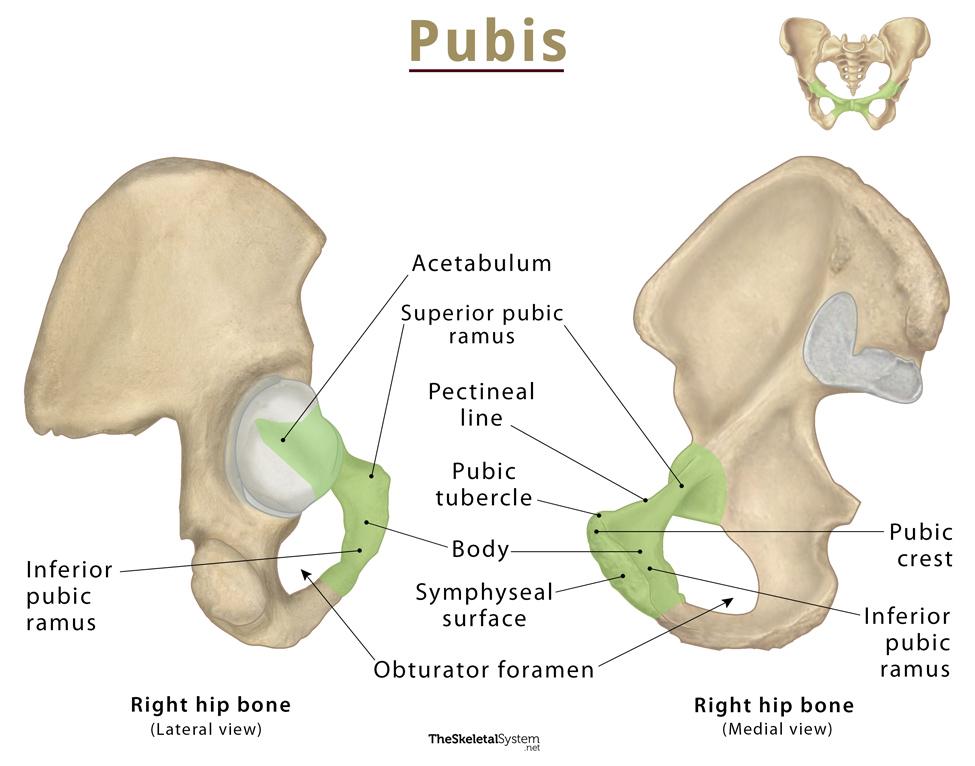
Pubis (Pubic Bone)
Pubis, also known as pubic bone, is the smallest component of the hip bone, forming its anterior and inferior parts. Its two main parts are the body and two rami (superior ramus and inferior ramus). The body is located anteromedially, and the two rami extend posterollatery from the body. The pubic body of the left and right hip bones articulates with each other at the pubic symphysis.
Articulations
1. Sacroiliac joint: It is a synovial joint formed between the ilium and the sacrum.
2. Pubic symphysis: The pubic bodies of the left and right hip bones articulate with each other, forming this secondary cartilaginous joint.
3. Hip joint: This ball-and-socket joint is formed between the hip bone and femur. The ball-like head of the femur fits into the socket-like acetabulum.
Muscle Attachments
Several muscles that attach to or originate from the hip bone are as follows:
Muscles that attach to the hip bone
1. Abdominal muscles:
- Abdominal external oblique muscle
- Abdominal internal oblique muscle
- Transversus abdominis muscle
2. Back muscles
- Multifidus muscle
3. Shoulder muscles
- Latissimus dorsi
Muscles that originate from the hip bone
1. Gluteal muscles
- Gluteus maximus muscle
- Gluteus medius muscle
- Gluteus minimus muscle
2. Lateral rotator group
- Piriformis muscle
- Superior gemellus muscle
- Obturator internus muscle
- Inferior gemellus muscle
- Obturator externus muscle
3. Hamstrings
- Long head biceps femoris
- Semitendinosus
- Semimembranosus
4. Anterior compartment of thigh
- Rectus femoris muscle
- Sartorius muscle
Ossification
The hip bone is ossified from eight centers: three primary and five secondary. The three primary centers give rise to the ilium, ischium, and pubis. The remaining five secondary ossification centers form the iliac crest, the inferior anterior spine, tuberosity of the ischium, the pubic symphysis, and one or more for the Y-shaped piece at the bottom of the acetabulum.
These ossification centers appear in the following order:
- For ilium, at the 8th week of fetal life.
- For ischium, during 4-6 months of gestational period.
- For pubis, during 4-6 months of fetal life.
- For acetabulum, during puberty.
The three bones, ilium, ischium, and pubis, fuse at 7-9 years via triradiate cartilage. The complete fusion, through the formation of the acetabulum, occurs at 20-25 years by replacing the triradiate cartilage.
References
- The Hip Bone – Teachmeanatomy.info
- Hip bone – Kenhub.com
- Hip bone – Anatomystandard.com
- Hip Bone Anatomy – Getbodysmart.com
- Innominate bones – Radiopaedia.org