Atlas Bone (C1)
Table of Contents:
Published on December 1st 2022 by staff
What is the Atlas
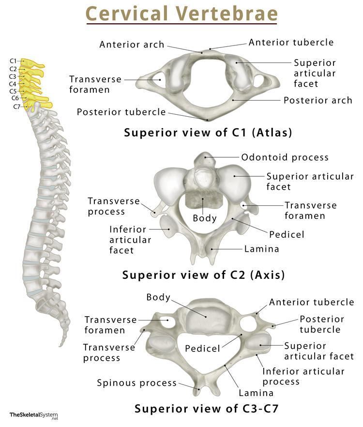
The human vertebral or spinal column can be divided into the cervical, thoracic, and lumbar spines. The cervical spine includes the first 7 vertebrae following the skull down the upper back. The atlas is the first of these 7 cervical vertebrae and is also referred to as the 1st cervical vertebrae or C1.
Its name derives from the Greek God Atlas, famous in mythologies for being condemned to bear the weight of the Earth on his shoulders for eternity. Similarly, the atlas supports the weight of the skull or the globe-like cranium.
Where is the Atlas Located
As mentioned above, the atlas is the first vertebral bone, located between the skull base and the axis (C2), the second cervical vertebrae.
Quick Facts
| Type | Irregular, atypical vertebra |
| How many are there | 1 |
| Articulates with | Axis (C1), occipital condyle |
Function
As it is the first bone of the vertebral column, the atlas is the point of connection between the skull and the spine. In addition to supporting the weight of the skull, the small bone also controls the head’s range of motion. The atlas forms a number of articulations and serves as the point of attachment for several muscles and ligaments that are instrumental to all head movements.
Anatomy
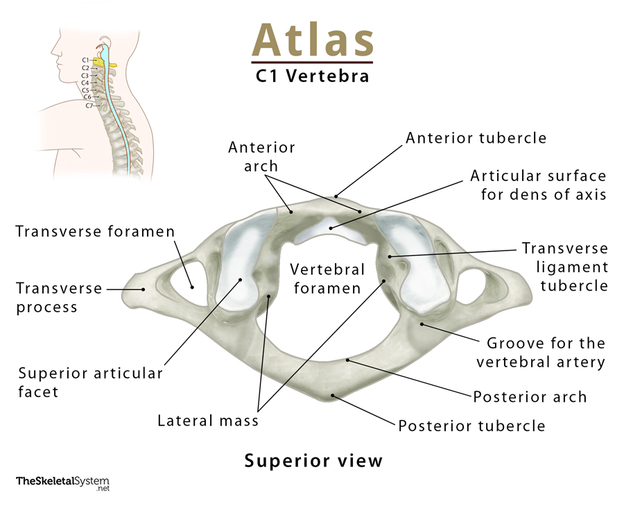
The atlas is a thin ring-shaped bone that does not resemble the typical vertebrae, thus being classified as an atypical vertebra. It is the most delicate of all the 7 cervical vertebrae and lacks a vertebral body and spinous process. Instead, it comprises an anterior and posterior arch and a lateral mass.
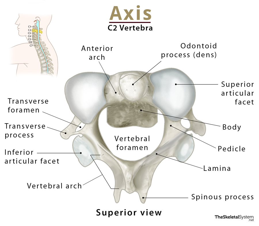
Anterior Arch
It is the bony band that forms the anterior part of the atlas’s ring. There is a bony prominence or tubercle at the front of the bone, known as the anterior tubercle, where the anterior longitudinal ligament attaches.
The smooth circular facet on its posterior surface articulates with the dens or odontoid process of the axis, forming the median atlantoaxial joint. It is a pivot joint that allows the rotational movements of the head, such as when you shake your head to say ‘no.’
The anterior atlanto-occipital membrane attaches to its upper border. In contrast, the lower border is the attachment point for the anterior atlantoaxial membrane.
Posterior Arch
As the name suggests, it is the posterior three-fourth part of the ring of the atlas that encloses the vertebral foramen. The thin band of bone has a slightly wider prominence at its apex in the back, known as the posterior tubercle. This bony landmark is similar in structure and function to the spinous process found in all the typical vertebrae. So, one of its primary purposes is to act as the attachment point for the nuchal ligament and the muscle rectus capitis posterior minor.
On both sides of the tubercle, on the superior surface of the posterior arch, are two marked depressions or grooves (grooves for the vertebral artery) to allow passage to the C1 spinal nerve and the vertebral artery.
The upper border of the posterior arch provides attachment to the atlanto-occipital membrane, while the lower border is where the posterior atlantoaxial membrane and the first pair of ligamentum flavum attach.
Lateral Mass
On either side of the anterior mass, along the inner surface of the ring, the bone thickens to form the two sturdiest regions of the atlas, the lateral masses. This is the part of the atlas that primarily supports the weight of the skull.
There is a smooth, kidney-shaped depression or facet on the upper surface (superior articular facet) of each lateral mass where the bone articulates with the condyles of the skull bone occipital to form the atlanto-occipital joint. This joint allows you to move your head to extend and flex the neck.
There is a short cylindrical projection on the lower surface of the mass called the inferior articular facet. These are incredibly smooth, articulating with the axis bone or C2 to form the lateral atlantoaxial joint. Along with the medial atlantoaxial joint, it also helps with the rotational movements of the head.
The medial surface of the lateral mass has another tubercle, the transverse ligament tubercle, which is the point of attachment for the transverse ligament.
Transverse Process
Each lateral mass has an irregular yet prominent ring-shaped lateral projection known as the transverse process. Each of these processes encircles a tiny opening called the transverse foramen, the passage for the vertebral artery and vein to pass through the neck. Apart from this, they also provide points of attachment to several muscles that aid in neck movement.
The transverse processes of the atlas and the posterior tubercle of the typical cervical vertebrae are homologous.
References
- C1 (Atlas): 1st Cervical Vertebra: InnerBody.com
- Atlas (C1):RadioPaedia.org
- Atlas Bone Anatomy: GetBodySmart.com
- Atlas: KenHub.com
- Atlas: HealthLine.com
- Cervical Spine Anatomy: UMMS.org