Axis Bone (C2)
Table of Contents:
Published on December 7th 2022 by staff
What is the Axis
The axis is 1 of the only 2 vertebrae with a unique name and purpose. It is the 2nd bone out of the 7 in the uppermost section of the spinal column, the cervical vertebra. It is also referred to as the C2 vertebra or epistropheus (rarely).
Its name derives from the fact that the odontoid process of C2 acts as the axis around which the C1 rotates to allow us the rotating movement of the head.
Where is the Axis Located
It is located in the superior part of the cervical spine, beneath the atlas or C1 vertebra, and above the third cervical vertebra or C3.
Quick Facts
| Type | Irregular, atypical vertebra |
| How many are there | 1 |
| Articulates with | Atlas (C1), third cervical vertebra (C3) |
Function
Since it is the second bone in the vertebral column, it plays a significant role in supporting the atlas and forming the atlantoaxial socket joint. So, along with the atlas, the axis controls and helps with all head and neck movements, keeping them flexible.
Anatomy
The basic structure of the bone can be divided into two parts — the anterior and posterior. All the bony markings and structures are grouped as the anterior and posterior components based on their location.
Filename: Axis Bone C2
Anterior Components
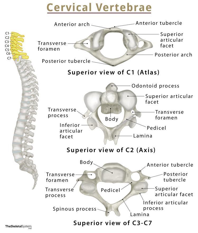
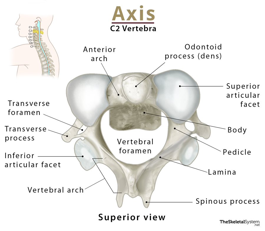
1. Odontoid Process (Dens)
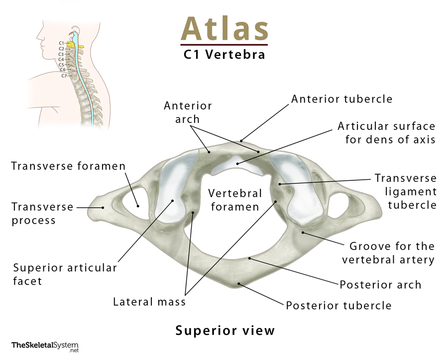
It is the most prominent and recognizable landmark on the anterior arch of the C2 vertebra. It is named after its tooth-like shape as ‘dens’ literally means the ‘tooth of the axis.’
The sturdy process projects superiorly to stick out through the vertebral foramen of the atlas bone above. Its smooth anterior articular facet articulates with the atlas’s anterior arch to form the median atlantoaxial joint. It is a pivot joint that allows for all the rotational movements of the neck. The atlas pivots around the knob-like odontoid process when you rotate your head to say ‘no.’
The transverse ligament of the atlas helps keep the dens axis in place. On both sides of the dens axis is a roughened region where the alar ligaments attach. It also has a pointy marking at its apex where the apical ligament attaches. These two ligaments connect the dens axis to the occipital condyles and foramen magnum.
2. Vertebral Body
The body is a thick cylindrical bony mass at the front of the axis, below the odontoid process. A longitudinal ridge at the front aspect of the body separates the two attachment points of the muscle longus colli. The body articulates with the body of the C3 vertebra below.
3. Superior Articular Facet
The two superior articular facets extend from the superior part of the bone’s body lateral to the odontoid process. The smooth, convex facet articulates with the cylindrical projecting inferior articular facet of the atlas, foming the lateral atlantoaxial joint.
At this joint, the two lateral masses of the atlas glide in a way so that one mass glides forward while the other glides backward. It allows the anterior arch of the atlas to rotate or pivot around the dens axis for the head to move in different directions.
4. Inferior Articular Facet
Like the superior articular facet, the two inferior articular facets extend from the inferior surface of the vertebral body to articulate with the C3 vertebra’s superior articular facet (C2-C3 uncovertebral joint).
5. Transverse Processes
These are the two small lateral projections extending sidelong upward from below the superior articular facet. The two ends of the transverse processes are marked with two tiny tubercles, where there is a small perforation known as the transverse foramen. The vertebral artery and vein pass through these two foramina.
These processes also provide attachment points to various head and neck muscles.
The rounded apex or tip and the posterior tubercle of typical cervical vertebrae are homologous.
6. Pedicles
The broad, sturdy lateral bony extensions from both sides of the body are called the pedicles. They slope towards the superior articular facet, extending laterally to the transverse processes.
A deep groove marks each pedicle at the front to let the vertebral venous plexus, vertebral artery, and suboccipital nerve pass. On the inferior surface, the Inferior vertebral notch holds the root sheath of the cervical spinal nerve 3 (C3).
Posterior Components
1. Laminae
The laminae extend medially from the transverse process towards the back. Projecting thick and strong from both sides, they meet at the posterior side of the bone, forming the vertebral arch. This forms the central opening or vertebral foramen at the center of the axis, allowing the spinal cord to pass. The vertebral arch takes care of one of the most important functions of the bone, which is keeping the spinal cord safe.
The vertebral foramen of the axis is smaller than that of the atlas.
2. Spinous Process
There is yet another narrow projection at the back of the axis from the point where the two laminae meet. It is called the spinous process. It provides a point for attachment for several muscles and ligaments that help move the head.
References
- Axis Bone Anatomy: GetBodySmart.com
- Axis: KenHub.com
- Axis: HealthLine.com
- Axis (C2): RadioPaedia.org
- The Cervical Spine: TeachMeAnatomy.info
- Axis: AnatomyStandard.com