Sacrum
Published on December 27th 2021 by staff
What is the Sacrum
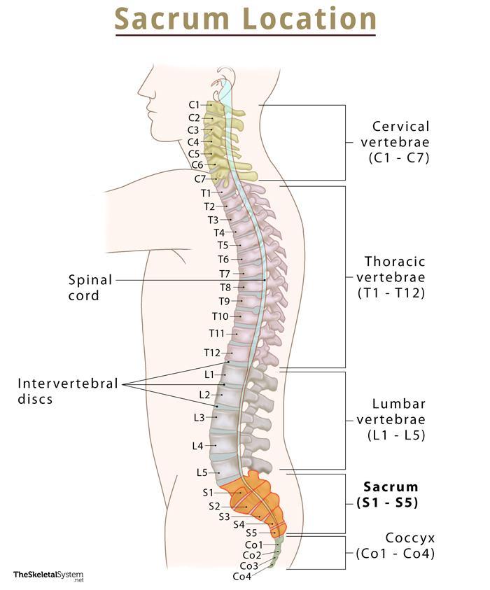
The sacrum is a large, flat, triangular-shaped, irregular bone, alternatively known as the sacral vertebra or sacral spine. It comprises five fused vertebrae (S1-S5), located at the base of the vertebral column or spine. The bone links the spine with the hip, thus helping in hip stability.
Where is the Sacrum Bone Located
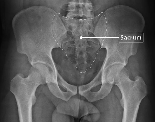
As stated, the sacrum bone is positioned at the base of the vertebral column or spine. More specifically, it is located between the right and left iliac bones of the hip and below the last lumbar vertebra (L5).
Quick Facts
| Type | Irregular bone |
| How many are there in the human body | 1 |
| Articulates with | Last lumbar vertebra (L5), coccyx or tailbone, and illium portion of the hip bone on both side |
Functions
- Locks the hip bones together on the posterior side, thus supporting the base of the spine.
- Surrounds and protects the spinal nerves of the lower back.
- Along with the hip bone, it forms the pelvic cavity, which supports and protects the delicate organs of excretory and reproductive systems.
- Aids in supporting and transmitting the body weight while standing or sitting.
- Provides stability of the hip.
Parts and Anatomy
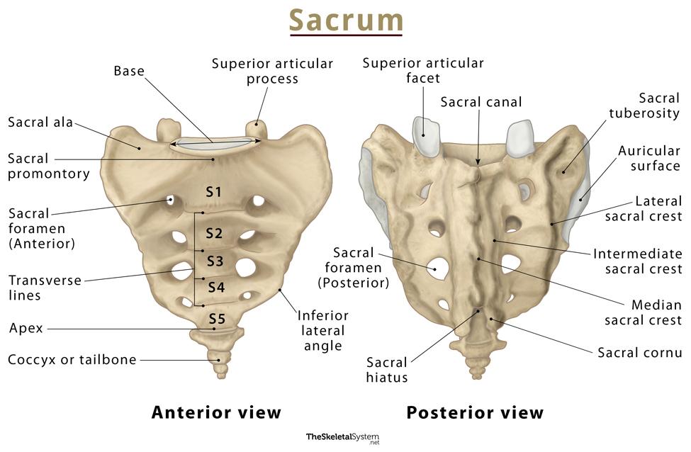
The sacrum is a concave, irregular bone, resembling an inverted triangle. The widest part, called the base, is at the top, and the pointy end, called the apex, is at the bottom. It also has three surfaces – dorsal, lateral, and pelvic. Along with these structures, the bone also has a hollow canal called the sacral canal that runs along its core.
Each part features several important bony landmarks.
Bony Landmarks
Base
The top part of the sacrum, lying just below the spinal base, is referred to as the base of the scarum. It is the widest portion of the bone. The first one of the five fused sacral vertebrae, S1, is located here. The S1 vertebra is the biggest one, having concave superior articular facets that project posteromedially to articulate with the fifth lumbar vertebra (L5). Both sides of the base bear a large wing-like projection known as an ala of sacrum or sacral ala. These alae articulate with the ilium bones of the pelvis, forming sacroiliac (SI) joints.
Apex
It is the pointy part of the sacrum, directing downwards. The fifth sacral vertebra lies in this most inferior segment of the bone. The apex projects posteriorly to increase the size of the pelvic cavity. This region features an oval facet for articulation with the coccyx.
Sacral Canal
The sacral canal is a hollow space that runs from the base to the apex of the sacrum. Internally, it is a continuation of the vertebral canal that runs along the core of the sacrum and ends at the fourth sacral foramina, as the sacral hiatus.
Though it is a continuation of the vertebral column, it does not contain the spinal cord, as the cord ends around the second lumbar vertebrae (L2). Instead, it has the cauda equina, which is a bundle of spinal nerve roots, and the filum terminale, a fibrous band of connective tissue.
Dorsal Surface
The dorsal surface of the sacrum is coarse and rugged due to the fusion of the sacral vertebrae, which gives rise to three bony ridges or crests, median, intermediate, and lateral.
The five sacral vertebrae fuse on the midline of the dorsal surface, giving rise to a central ridge called the median sacral crest. It is formed by the fusion of the spinous processes of the first three sacral vertebrae. The supraspinous ligament attaches here.
All the articular processes of the sacrum, except the superior articulating process of S1 and the inferior articulating process of S5, fuse to give rise to the intermediate sacral crests. The posterior sacroiliac ligaments are attached along this crest. The top part of S1 articulates with the inferior articulating process of L5, while the bottom part of S5, known as the sacral cornu, articulates with the coccyx.
The transverse processes of the five sacral bones fuse to form the lateral sacral crests, which serve as a point of attachment for the posterior sacroiliac and sacrotuberous ligament. The processes do not fuse completely, thus leaving four pairs of openings on either side, called posterior sacral foramina. The sacral nerve fibers enter and leave the sacral canal through these four pairs of sacral foramina.
Lateral Surface
The lateral surface of this bone is broad above but narrows into a thin edge as it travels down. The upper part of the surface presents an ear-shaped area, the auricular surface, which remains covered with cartilage, and articulates with the ilium. There is a rough prominence behind the auricular surface called the sacral tuberosity. It contains three uneven depressions for the attachment of the posterior sacroiliac ligament. The sacrotuberous and sacrospinous ligaments, along with some fibers of the gluteus maximus, get attached to the lower half of the lateral surface.
Pelvic Surface
The pelvic surface of the sacrum is relatively smoother than its dorsal surface. The surface is marked by four transverse lines, which are the remnants of the fused sacral intervertebral discs.
Superiorly, there is an anterior bony projection, known as the sacral promontory. It marks the posterior margin of the pelvic inlet and is continuous with the margin of the sacral ala. The front sides of the four pairs of sacral foramina are present on this surface.
Articulations
- Lumbosacral joint: The base of the sacrum articulates with the fifth lumbar vertebrae (L5) superiorly via the L5/S1 intervertebral disc, forming this amphiarthrodial joint.
- Sacrococcygeal joint: Here, the apex of the bone articulates with the base of the coccyx, to form another amphiarthrodial joint.
- Sacroiliac joint: The sacral ala laterally articulates with the ilium of the pelvis, forming this synovial joint.
Ossification
Though we know the sacrum is a single bone, it does not look like this from the beginning. Humans are born with 4-6 sacral vertebrae instead of a single bone. As one grows up, these vertebrae fuse to form a single bone, and the overall shape of the sacrum solidifies. The fusion does not take place in all sacral vertebrae simultaneously. It begins with the fusion of S1 and S2.
The process typically starts in the mid-teens and gets completed in the early to mid-twenties. This ossification process is thought to start earlier in females than males.
Anatomical Variations of the Sacrum
Sometimes, the sacrum shows some anatomical variation, including variation in the number of vertebrae, its surface and curvature.
- The most common anatomical variation of the bone is the variation in the number of sacral vertebrae. Commonly sacrum has five fused vertebrae, but four or six sacral vertebrae have also been documented.
- Another anomaly of the sacrum is related to its surface and curvature. The curvature of the bone varies widely between individuals.
- In some individuals, the first and second sacral vertebrae do not fuse. Instead, they remain separately articulated.
Sacrum in Females vs. in Males
Sacrum is sexually dimorphic, meaning it has a slightly different appearance in females and males. The sacrum is wider in females than males. It is also more backwardly curved in females, increasing the size of the pelvic cavity. This wider pelvic cavity in females aids in enduring pregnancy, offers more space for the developing fetus, and houses reproductive organs.
Muscle Attachments
Several lower limb and back muscles originate or get inserted on the sacrum.
Originating from the sacrum
- Piriformis
- Iliacus
- Multifidus lumborum
Inserting on the sacrum
- Coccygeus
- Erector spinae
Nerves
As mentioned, the cauda equina, long sacral roots of spinal nerves, pass through the sacrum via the sacral canal.
These nerves enter the sacrum from the vertebral foramen of the lumbar vertebrae through the sacral canal. From there, they branch out and exit the bone through four pairs of sacral foramina or the sacral hiatus, present at the bottom end of the canal.
FAQs
Q.1. Is the sacrum part of the axial skeleton or the appendicular skeleton?
Ans. The sacrum is the part of the axial skeleton.
References
- Sacrum – Kenhub.com
- The Sacrum – Teachmeanatomy.info
- Sacrum – Innerbody.com
- Sacrum – Radiopaedia.org
- Anatomy, Back, Sacral Vertebrae – Ncbi.nlm.nih.gov