Orbital Bones
What is the Bony Orbit
The bony orbits or orbital cavities, are the two symmetrical, bilateral cavities in the skull that surround and protect the eyeballs and other soft tissues in the region. They have a squarish pyramid shape resembling a pear.
Names of the Bones of the Orbit With Basic Anatomy
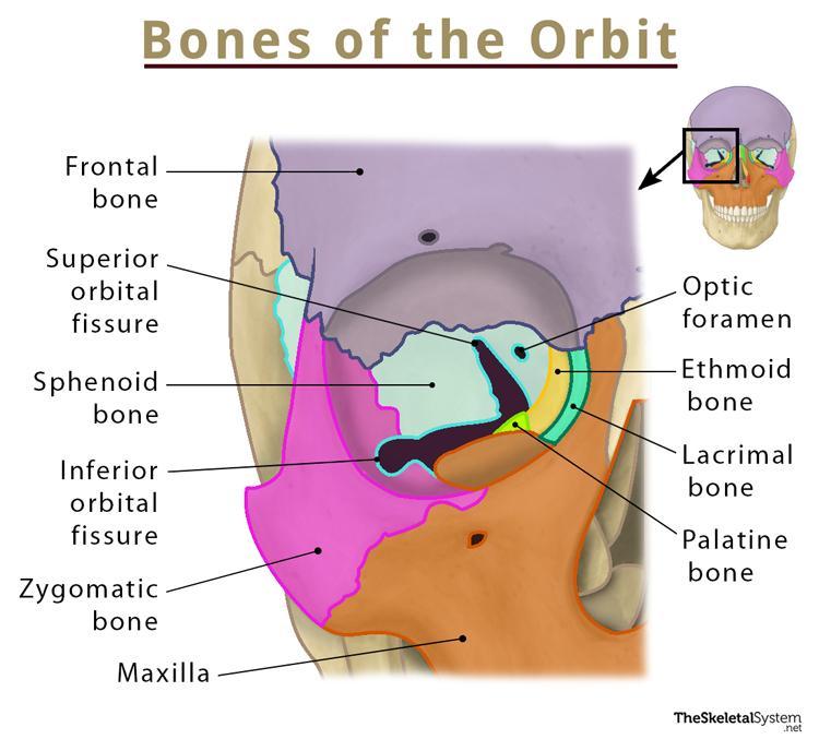
7 of the cranial and facial bones contribute to the formation of the orbital cavities, with 3 being cranial bones and the other 4 being facial bones:
- Sphenoid (cranial)
- Frontal (cranial)
- Ethmoid (cranial)
- Zygomatic (facial)
- Lacrimal (facial)
- Maxilla (facial)
- Palatine (facial)
Diagram – Bones of the Orbit
An easy way to remember the names of these bones is a memorizing this simple mnemonic:
Many Friendly Zebras Enjoy Lazy Summer Picnic
M – Maxilla
F – Frontal
Z – Zygomatic
E – Ethmoid
L – Lacrimal
S – Sphenoid
P – Palatine
These bones make up the walls of the bony orbits.
Walls and Surfaces With Important Landmarks
As mentioned above, the orbits are like a pair of pear-shaped caverns with six sides or surfaces:
1. Base
Also called the orbital margin or rim, it is the side of the orbit that opens toward the front of the face. It is bordered by the eyelids and has the following 4 margins:
- Supra-orbital: Formed by the frontal bone
- Medial: Formed by the frontal process of the maxilla
- Infra-orbital: Formed by the zygomatic bone and maxilla’s zygomatic process
- Lateral: Formed by the zygomatic bone and its frontal process, along with the frontal bone’s zygomatic process
2. Apex
It is the posteromedial surface pointing towards the skull. This side of the cavity is bound by the sphenoid bone. This is where the optic foramen opens up into the optic canal. The optic nerve (CN II — plays a vital role in vision) and the ophthalmic artery pass through the optic foramen to exit the skull.
3. Roof
It is the superior wall of the orbital cavity, bound by the frontal bone and the sphenoid’s lesser wing. This is the wall that marks the division between the orbital cavity and the anterior cranial fossa. The lacrimal fossa, a vital skull landmark, is located here. It houses the orbital lobe of the lacrimal gland, which plays a vital role in tear secretion.
4. Medial Wall
As the name suggests, this is located medially or towards the side of the nose. 4 bones contribute to forming this wall – ethmoid, lacrimal, maxilla, and sphenoid.
The orbital plate of the ethmoid bone, a rather thin portion of the bone also called ‘lamina papyracea’, is the primary contributor to this wall. The lacrimal bone, along with the maxilla’s frontal process, forms the lacrimal groove, which houses the lacrimal sac. Behind the ethmoid, the sphenoid’s lesser wing forms the part of the optic foramen located on this wall.
This wall separates the bony orbits from the ethmoid sinuses – the hollow spaces around the nasal bones.
Apart from the lacrimal groove, this wall has three other important landmarks – the anterior and posterior ethmoidal foramina and the only cartilaginous portion in the orbit, the trochlea.
5. Floor
It is the inferior wall formed primarily by the maxilla’s orbital surface, with the zygomatic, and palatine bones making up the rest. It acts as the separator between the orbital cavity and maxillary sinus, another pair of hollow spaces around the nasal bones.
The inferior orbital fissure, which allows passage to a number of neurovascular structures, is the most important landmark on the bony orbit floor.
6. Lateral Wall
This is the outer wall on the side of the ear, with its anterior side formed by the zygomatic bone, while the sphenoid’s greater wing forms the posterior side. Being the strongest and thickest part of the orbit, it plays the wall between the orbit, and the temporal and middle cranial fossae.
The superior orbital fissure is located here. Being one of the primary pathways to enter or exit the orbital cavity, it allows passage to the zygomatic nerve, the sympathetic nerves, and the inferior ophthalmic vein.
Contents Of the Bony Orbits
- Most of the space within these cavities is occupied by the globe or eyeballs.
- The rest of the hollow space contains orbital fat that keeps the eye and its muscles in place.
- The extraocular muscles attach to the eyeballs allowing us to move our eyes and the upper eyelids.
- The cranial nerves that innervate the eyes are also among the contents of the optical cavities. These include the optic, trochlear, oculomotor, abducens, and trigeminal nerves.
- The lacrimal apparatus controlling the production and release of tears is also located here.
- The rest of the components that fill the remaining space are blood vessels, including branches of the ophthalmic artery and the inferior and superior ophthalmic veins
References
- Bones of the orbit – Kenhub.com
- The Bony Orbit – Teachmeanatomy.info
- Anatomy, Head and Neck, Orbit Bones – Ncbi.nlm.nih.gov
- Anatomy of the orbit – Osmosis.org
- Bones of the Orbit – Springer.com