Frontal Bone
Published on March 1st 2022 by staff
What is the Frontal Bone
The frontal bone is one of the eight cranial bones. In Latin, ‘frons’ stands for ‘forehead’, which gives the bone its name as it forms the smooth curve of the forehead. Along with the forehead, the bone partially forms the bony part of the nose and the upper ridge and roof of the eye orbit. This unpaired bone supports and protects the brain’s delicate nervous tissues, supports several head muscles, and contributes to the characteristic shape of the skull.
Where is the Frontal Bone Located
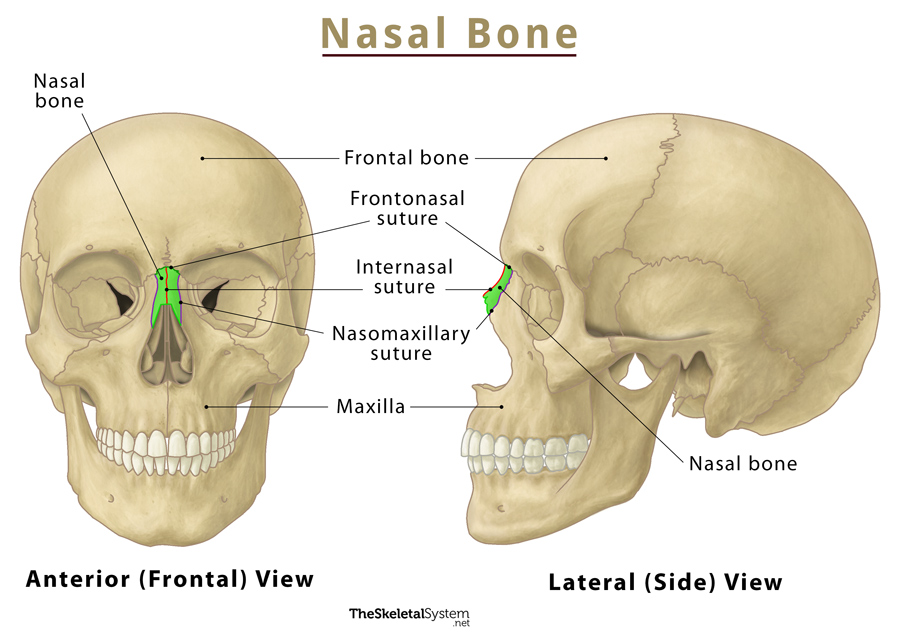
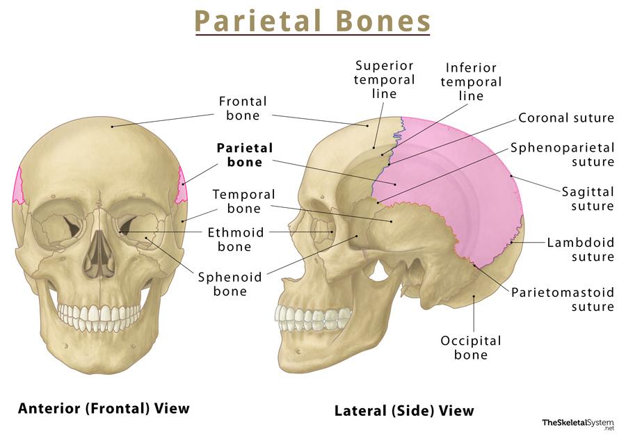
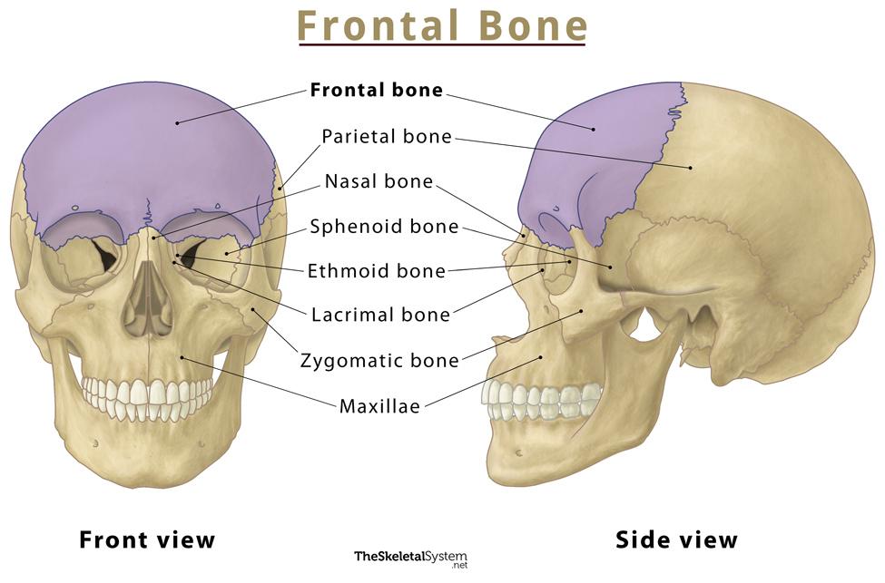
The frontal bone is located at the front of the skull, above the nasal bones, and in front of the parietal bones. You can easily feel the bone by touching your forehead.
Quick Facts
| Type | Flat bone |
| How many are there in the human body | 1 |
| Articulates with | 12 bones: sphenoid, ethmoid, parietal (2), nasal (2), maxillae (2), lacrimal (2), and zygomatic (2) bones |
Functions
- Protects the frontal lobe of the brain.
- Supports the structures of the head, such as the eye orbits and nasal passages.
- Aids facial expression through its muscle attachments.
- Forms the forehead and plays a major role in a person’s appearance.
Frontal Bone Anatomy
It is a bowl-shaped bone comprising three parts: squamous, orbital, and nasal.
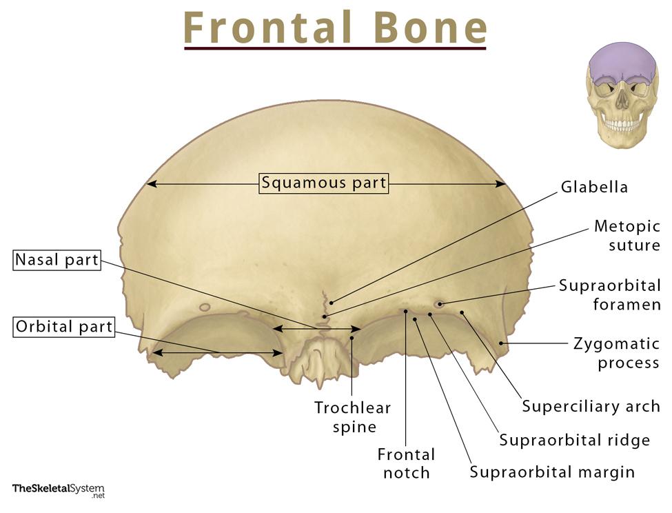
1. Squamous Part
It is the largest area of the bone, encircling the forehead. Its external side is flat, but the internal side is concave. Two thickened regions, the supraorbital notches or supraorbital ridges, form the characteristic shape of the two brows and the anterior cover for the frontal sinuses.
Deep into the supraorbital ridges are a pair of hollow spaces known as the frontal sinuses. These sinuses connect to the nasal cavity and are lined with mucous membranes. Though their exact function is unknown, the hollow spaces are believed to make the skull lighter and improve vocal tone by increasing the resonance of the skull.
Above each orbit in the supraorbital ridge, there are two supraorbital foramina. The supraorbital nerve and vessels exit through these foramina. The inferior end of each supraorbital ridge features the supraorbital margin. Here, the bone forms a sharp angle to form the superior and medial margins of the eye orbits. Inside the orbits, the bone continues posteriorly along the superior margin until it meets the sphenoid bone. It also continues inferiorly along the medial margin until it joins the ethmoid and lacrimal bones.
The arches above the supraorbital notches are known as the superciliary arches. Medial to the supraorbital ridges, a small, smooth, slightly elevated surface called glabella is present that ends inferiorly at the nasal bones.
It also contains a small zygomatic process that arises caudolaterally. The bone articulates with the zygomatic bone via this process.
2. Orbital Part
This part of the bone forms the roof of the eye orbit and the ethmoidal sinuses, lying between the eyes and nose. The orbital part consists of two orbital plates separated by a gap known as the ethmoidal notch. The ethmoid air cells lie within this notch. There are two openings at the front and back of this part, the anterior ethmoidal foramen and posterior ethmoidal foramen. These foramina allow the passage of anterior and posterior ethmoidal vessels and nerves, respectively. The orbital part also features another important bony landmark, called the trochlear spine or fovea trochlearis which serves as an insertion site for the superior oblique muscle.
3. Nasal Part
This part of the bone is present between the brow ridges, hence the name. It ends in a serrated nasal notch that articulates inferiorly with the nasal bones and laterally with the frontal processes of the maxilla and lacrimal bones. The stem of the nose is formed due to these articulations.
Borders and Articulations
As stated, the bone articulates with two unpaired (sphenoid, ethmoid) and five paired (parietal, nasal, maxilla, lacrimal, zygomatic) bones via sutures. Below is the list of the articulating bones, along with their respective sutures:
- Sphenofrontal suture: WIth the sphenoid bone
- Frontoethmoidal suture: With the ethmoid bone
- Coronal suture: With the parietal bones
- Frontonasal suture: With the Nasal bones
- Frontomaxillary suture: With the maxilla
- frontolacrimal suture: with the lacrimal bones
- Zygomaticofrontal suture: With the zygomatic bones
Muscle Attachments
The temporalis and orbicularis oculi are two major facial muscles originating from the frontal bone. Another one, called the frontalis muscle, which forms the frontal belly of the epicranius muscle, passes over the smooth squamous part of the bone.
Ossification and Development
Like other skull bones, frontal bone is derived from neural crest cells. The bone evolves as two separate parts at birth, joined by a frontal suture. The whole frontal suture undergoes intramembranous ossification at two years, except its anteriormost part, which ossifies around eight years of age, but may even persist throughout life. This remnant suture is termed the metopic suture.
References
- Frontal bone – Kenhub.com
- Anatomy, Head and Neck, Frontal Bone – Ncbi.nlm.nih.gov
- Frontal bone – Radiopaedia.org
- Frontal Bone – Innerbody.com