Maxilla
Published on April 12th 2022 by staff
What is Maxilla
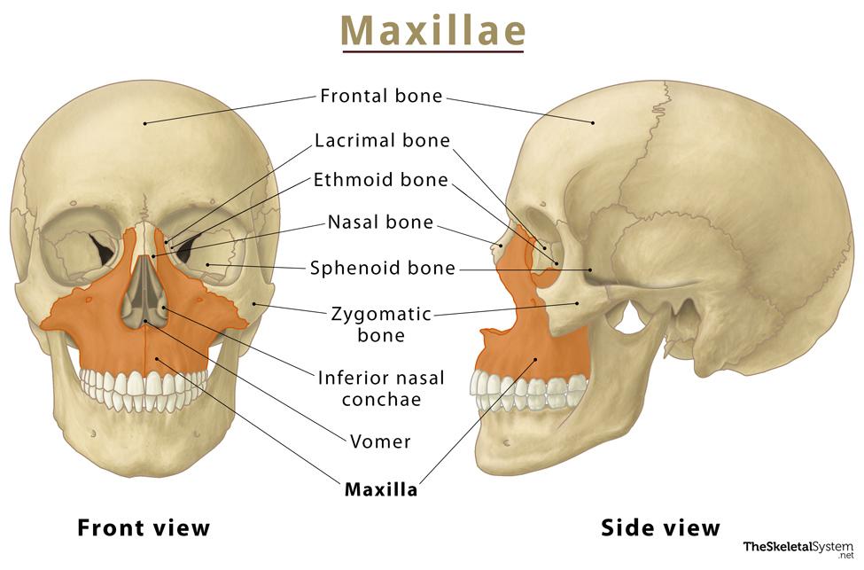
Maxilla (plural: maxillae) is one of the eight facial bones that form the facial skeleton. It is the second largest bone of the face. As it forms the upper jaw holding the upper set of teeth, it is sometimes referred to as the upper jaw bone. It also forms the lower parts of eye sockets and nasal cavities.
Though the maxilla looks like a single bone, it is a paired bone, joined by a delicate suture in the middle called an intermaxillary suture or median palatine.
Where is the Maxilla Located
The bone is positioned in the mid-face, just above the mandible. Starting from the middle of the forehead, it extends to the sides of the nose, reaching up to the cheekbones. You can easily feel the bone by pressing the skin under your cheekbones.
Quick Facts
| Type | Irregular Bone |
| How many are there in the human body | 2 |
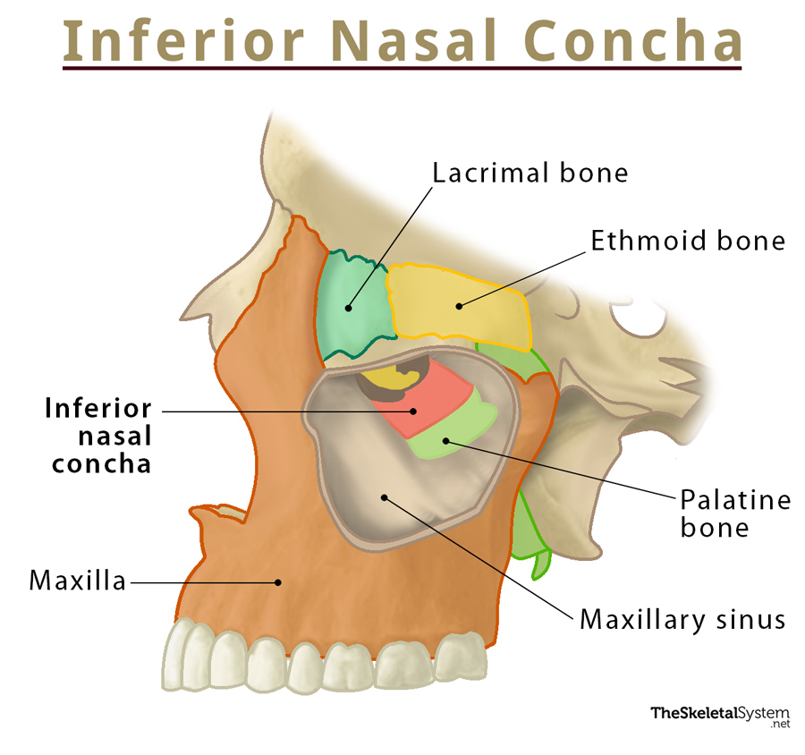
| Articulates with | 9 bones: frontal, ethmoid, sphenoid, nasal, zygomatic, lacrimal, inferior nasal concha, palatine, vomer. |
Functions
The left and right maxilla collectively help to:
- Hold the upper set of teeth in place.
- Increase the volume and depth of voice.
- Form a part of the eye socket, nasal cavity, and hard palate.
- Play an important role in chewing and communication.
Anatomy
The bone consists of a body and four projections or processes: frontal, zygomatic, palatine, and alveolar.
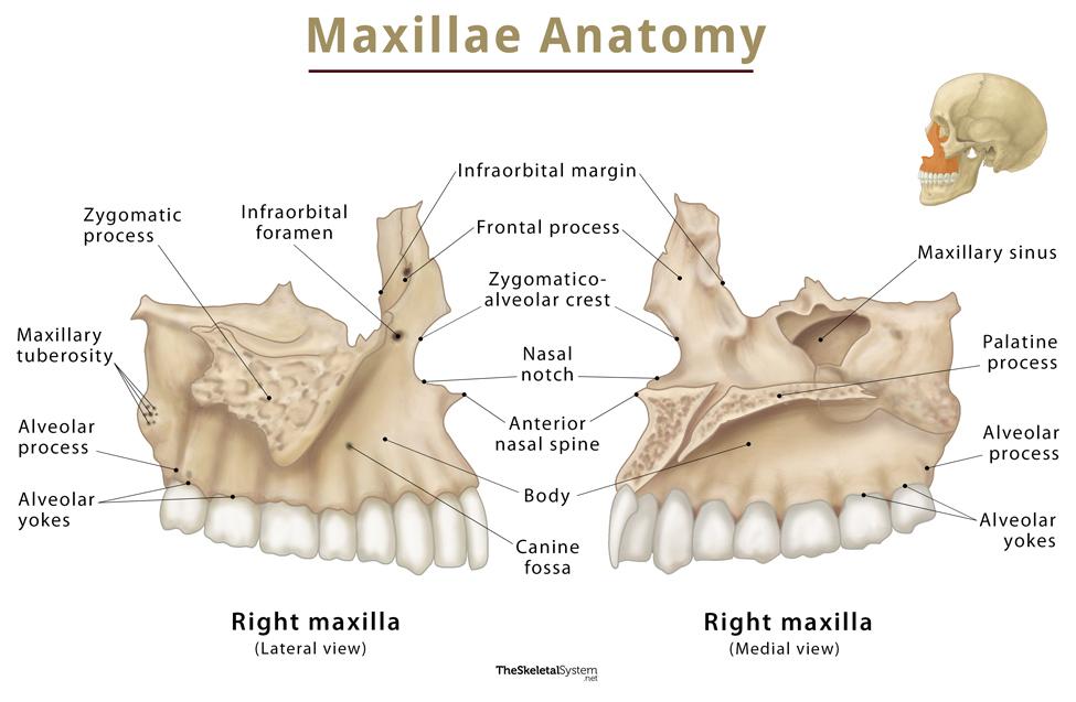
1. Body
It is the largest part of the bone, having a roughly pyramidal shape and four surfaces: anterior, orbital, nasal, and infratemporal surfaces.
i. Anterior surface
The anterior surface of the body features a curved margin of the bony anterior nasal aperture, called the nasal notch. This surface also partly forms the lower margin of the eye socket, named as infra-orbital margin, along with the zygomatic bone. Below the infra-orbital margin is an opening on the anterior aspect of the maxilla, the infra-orbital foramen. This foramen is the outer opening of the infra-orbital canal that serves as the passage for the infraorbital nerve and blood vessels. Just below the infra-orbital foramen, there is another depressed area on the anterior surface of the body called the canine fossa. The levator anguli oris muscle originates from here.
ii. Orbital surface
This surface forms most of the floor of the eye socket and features the infra-orbital groove leading into the infra-orbital canal. This canal ends with an opening on the anterior surface of the maxilla and serves as the passage for the infraorbital nerve and blood vessels.
iii. Nasal surface
The nasal surface of the body of the maxilla forms part of the lateral wall of the nasal cavity and features a large opening, the maxillary hiatus. The maxillary hiatus is an opening to the maxillary sinus, located below the middle nasal concha. The maxillary sinus is a paranasal sinus, an air-filled cavity located within the body of the maxilla.
iv. Infratemporal surface
The infratemporal surface of the body of the maxilla forms the anterior wall of the infratemporal fossa. It also features the maxillary tuberosity on the inferior aspect of lateral margin. The maxillary tuberosity or maxillary eminence has several small openings called alveolar foramina that lead into the alveolar canals. These canals transmit the posterior superior alveolar nerves and posterior superior alveolar arteries and veins to the upper teeth. A few fibers of the medial pterygoid muscle originate from this maxillary tuberosity.
2. Alveolar Process
It is a curved, horseshoe-shaped process located on the inferior surface of the maxilla. With its porous structure, it holds the teeth of the upper jaw. The process has a curved free margin called the alveolar arch, housing the dental alveoli (dental sockets), alveolar yokes, and the interalveolar and Interradicular septa. Channels in the alveolar process allow passage to the alveolar arteries, alveolar nerves, and the periodontal ligaments that innervate, irrigate, and keep the upper teeth in place.
3. Frontal Process
The frontal process is a bony extension that projects outward and upward to articulate with the frontal bone superiorly and the nasal bones medially. It forms the lower and central portions of the forehead along with the frontal and nasal bones. A vertical ridge on the frontal process forms the anterior lacrimal crest or the medial border of the bony orbit. It also forms the lacrimal groove posteriorly along with the lacrimal bone. It is also connected to the anterior ethmoidal sinuses on the superomedial side.
4. Zygomatic Process
The zygomatic process is the most lateral portion of the maxilla, growing laterally to meet the zygomatic bone. It forms the superolateral border of the maxillary sinus and is superior to the first maxillary molar. The process inferiorly shares a border with the alveolar process, while its superomedial border is shared with the frontal process. Along with the alveolar process, the zygomatic process also plays a crucial role in providing structure to the midface. On the anterior surface, lateral to the zygomatic process, a depression is formed known as the canine fossa, which constitutes the anterior surface of the maxilla. Another depression, the zygomaticoalveolar crest, is located below the zygomatic process and above the alveolar process.
5. Palatine Process
It is a horizontal plate that extends medially to form the oral cavity roof and nasal cavity floor. The palatine process of the maxilla, along with the palatine bone, forms the hard palate. There is an opening called the incisive foramen on the median line of the process behind the incisors that lets the greater palatine vessels and the nasopalatine nerve pass. The small process called the anterior nasal spine is also located here.
Articulations
As stated, each maxilla articulates with the following 9 bones:
- Frontal bone: Superiorly
- Sphenoid, palatine, lacrimal, and ethmoid bones: Posteriorly
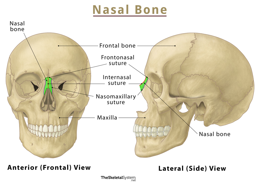
- Nasal bone and vomer: Medially
- Nasal concha: Inferiorly
- Zygomatic bone: Laterally
The left and right maxillae also articulate with each other, appearing like a single unpaired bone.
Muscle Attachments
Following are the muscles that attach to the maxillae.
Originating from the maxillae
- Levator labii superioris alaeque nasi
- Levator labii superioris
- Levator anguli oris
Inserting into the maxillae
- Zygomaticus minor
- Buccinator
- Orbicularis oris
Ossification and Development
All five parts of the maxilla undergo intramembranous ossification through two ossification centers.
References
- Maxilla — Kenhub.com
- Anatomy, Head and Neck, Maxilla — Ncbi.nlm.nih.gov
- Maxilla — Radiopaedia.org
- Maxilla — Sciencedirect.com