Ethmoid Bone
Table of Contents:
Published on April 23rd 2022 by staff
What is Ethmoid Bone
The ethmoid is a small unpaired cranial bone that separates the nasal cavity from the brain. The bone got its name from the Greek’ ethmos’, meaning sieve, due to its lightweight and spongy texture.
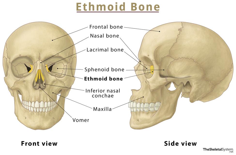
Where is the Ethmoid Bone Located
It is located in the center of the skull, specifically at the roof of the nose, between the two orbits.
Quick Facts
| Type | Irregular Bone |
| How many are there in the human body | 1 |
| Articulates with | Maxillae, inferior nasal conchae, vomer, nasal, frontal, lacrimal, palatine, and sphenoid bones. |
Function
It forms the walls of the eye socket, nasal cavity, nasal septum, and the anterior cranial fossa floor.
Anatomy
This cubical bone is made up of three parts – the cribriform plate, the perpendicular plate, and two ethmoidal labyrinths.
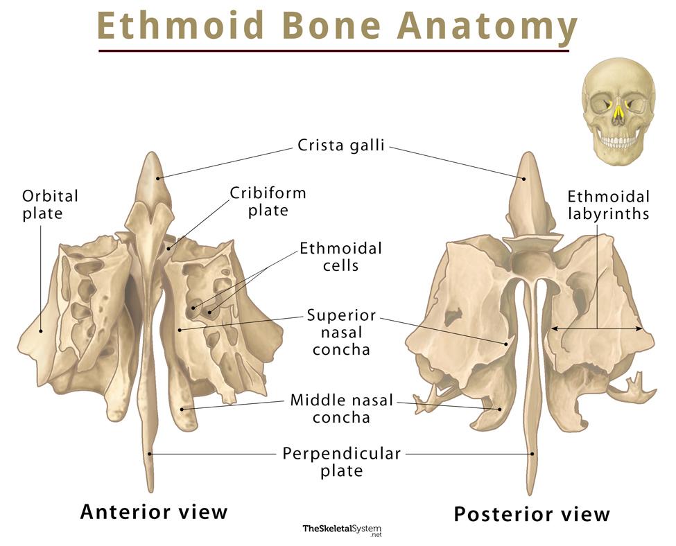
1. Cribriform plate
This part of the ethmoid bone lies within the frontal bone’s ethmoidal notch, forming the nasal cavity roof. The structure has derived its name from the Latin’ cribriform’, meaning ‘perforated’, as this plate has several perforations, giving it a sieve-like appearance. It allows passage to the olfactory fibers to reach the anterior cranial fossa from the nasal cavity. The plate also features a small, vertical bony projection called crista galli, where the falx cerebri (a crescent-shaped sheet of dura mater that separates the two cerebral hemispheres) is attached.
2. Perpendicular plate
It is a thin lamina descending from the cribriform plate, inferiorly attaching to the septal cartilage to form the superior two-thirds of the nasal septum.
3. Ethmoidal labyrinths
The ethmoid labyrinths are large masses located on both sides of the perpendicular plate. They are made up of multiple thin-walled compartments known as ethmoidal cells, which comprise the ethmoidal sinus. The number of these cells increases with age. These labyrinths also form a part of the superior and middle nasal concha.
Each labyrinth is composed of two sheets of bone – an orbital plate and a medial sheet. The orbital plate is the lateral sheet of bone that forms the orbit’s medial wall. On the other hand, the medial sheet makes up the upper lateral wall of the nasal cavity, from which the superior and middle nasal conchae descend into the nasal cavity.
The external edges of these labyrinths are referred to as the orbital lamina or lamina papyracea. This lamina connects with the frontal bone, palatine bone, lacrimal bone, and sphenoid bone, forming the major portion of the inside wall of the eye cavity.
Muscle Attachment
As ethmoid bone forms a part of the orbit, it is associated with the following seven extraocular muscles:
- Lateral rectus
- Medial rectus
- Superior rectus
- Inferior rectus
- Superior oblique
- Inferior oblique
Articulations
The ethmoid bone articulates with 5 paired and 3 unpaired bones:
- Paired bones – Inferior nasal conchae, maxillae, lacrimal, palatine, and nasal bones.
- Unpaired bones – Frontal, vomer, and sphenoid bones.
References
- Ethmoid bone — Kenhub.com
- The Ethmoid Bone — Teachmeanatomy.info
- Anatomy, Head and Neck, Ethmoid Bone — Ncbi.nlm.nih.gov
- Ethmoid bone — Radiopaedia.org
- Ethmoid bone — Anatomystandard.com