Ankle Bones
The ankle is the region in the human leg where the lower leg meets with the proximal end of the foot. The ankle allows us to move the feet in different directions.
Names and Anatomy of the Bones in the Ankle
Though often believed to contain just one joint, the ankle is actually formed of two joints – the ankle joint, also known as the tibiotalar or talocrural joint, and the subtalar joint. Here are the 4 bones that form the ankle:
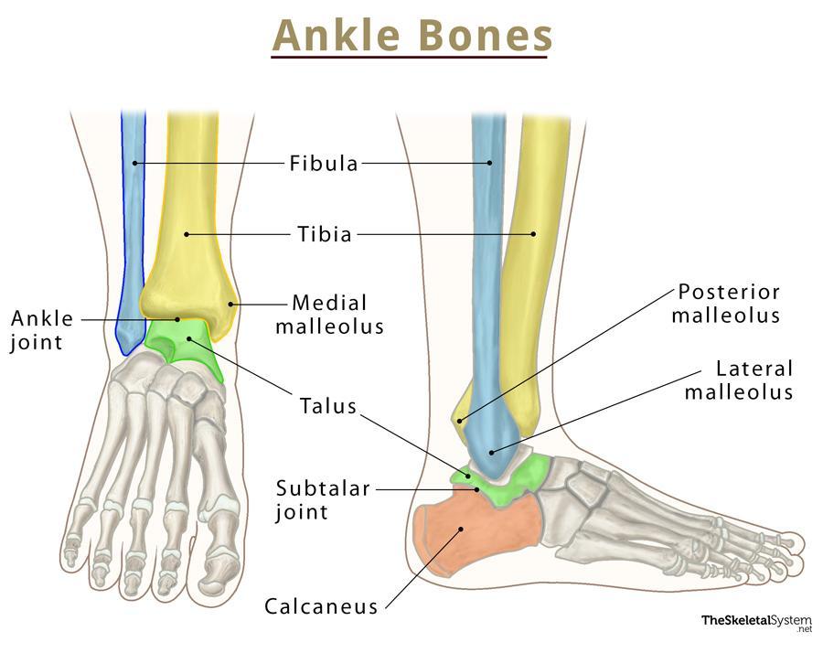
Ankle Joint: It is a synovial joint, more particularly a hinge joint between the tibia, fibula, and talus. This joint allows us to move the ankle up and down (plantarflexion and dorsiflexion).
Subtalar Joint: Another synovial joint, it is located below the ankle joint. The bones that articulate to form this joint are the talus and calcaneus.This joint helps with turning the foot to the left and right (inversion and eversion).
Bony Protrusions
There are a number of bony protrusions in the region that serve as attachment points for the ankle muscles and ligaments.
- The medial malleolus can be felt on the inside of the ankle; it is a part of the base of the tibia
- The posterior malleolus is a bony bump at the back of the ankle; This is also a part of the base of the tibia
- The lateral malleolus is the bump you can feel on the outside of the ankle; it is the bony protrusion on the lower end of the fibula
Muscles and Tendons
A number of muscles originating in the lower leg are responsible for all the movements of the ankle and foot. These include:
- Calf muscles
- Tibialis Posterior muscle
- Peroneal Brevis
- Peroneal Longus
The largest tendon in the human body, the Achilles tendon, is located in this region. It is where the calf muscles attach. Other important tendons here include:
- Flexor hallicus longus
- Flexor digitorum
- Anterior tibialis tendon
- Posterior tibialis tendon
- Peroneal tendons
Ligament Attachments
- Anterior and posterior talofibular ligament: Attaches the talus and fibula
- Calcaneofibular ligament: Runs from the fibula to the calcaneus
- Anterior tibiofibular ligament: Forms the connection between tibia and fibula.
- Lateral collateral ligaments: Forms the connection between fibula and calcaneus; stabilizes the outer part of the ankle.
- Deltoid ligaments: Runs from the tibia to talus, calcaneus, and navicular bone; stabilizes the inner part of the ankle.
References
- The Basics Of Ankle Anatomy And Foot Anatomy – Csog.net
- Ankle Anatomy – Arthritis.org
- Parts Of The Ankle; Ankle Bone Anatomy – Arlingtonortho.com
- Ankle – Image, Function, Conditions, & More – Webmd.com
- Foot and ankle bones – Mayoclinic.org