Fibula
What is the Fibula
Fibula is one of the two long bones found in the lower leg. The other bone found in the lower leg is tibia. The fibula is smaller and thinner than the tibia. It is commonly known as calf bone, running parallel to the tibia.
The name of the bone has a Latin origin, where fibula means ‘brooch’. According to popular belief, it is named so because when fibula pairs with the tibia, it looks like the safety pin of a brooch.
Where is the Fibula Located
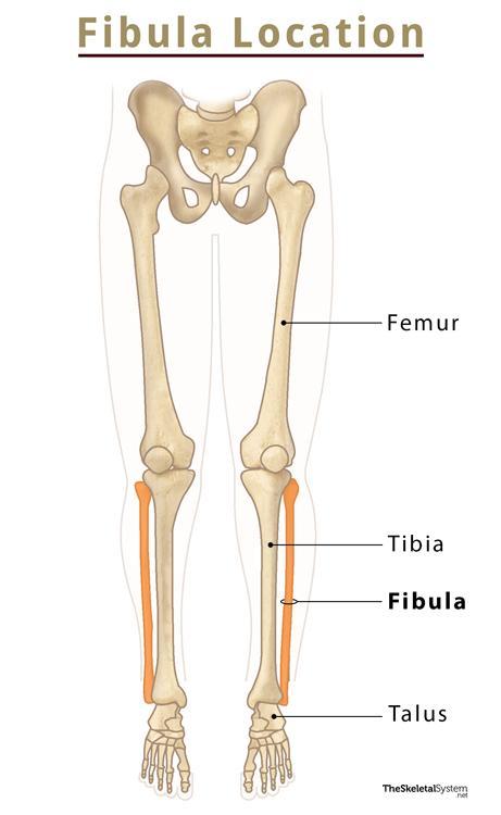
Fibula is located on the lateral side of the lower leg, just below the knee. It runs alongside the tibia, extending below the femur to the ankle.
Quick Facts
| Type | Long bone |
| Length | In adult males: Approximately 39 cm. In adult females: Approximately 36 cm. |
| How many are there in the human body | 2 (1 in each leg) |
| Articulates with | Tibia and talus |

Functions
- Provide stability to the lower limb and the ankle joint by combining with the tibia.
- Act as a lever during ankle movements, providing a range of motion during rotation of the ankle.
- Support the muscles of the lower leg.
Anatomy – Parts of the Fibula with its Bony Landmarks
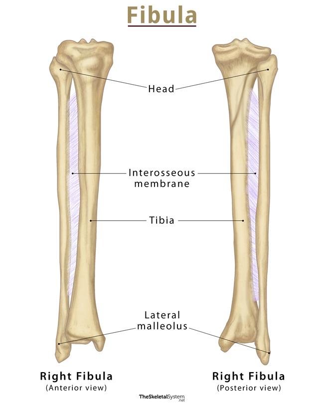
Like any other long bone, fibula also has two ends, proximal and distal parts, and an intervening shaft. Each of its parts features several important bony landmarks.
Proximal Part
The proximal end of the fibula features a slightly rounded enlargement known as its head. It bears a circular articular facet for articulation with the lateral condyle of the tibia. Lateral to the facet, there is an upward projection, known as the styloid process that extends superiorly from the head. Just below the fibular head, there is a short bare region referred to as the neck.
Shaft
The shaft or body makes up the major portion of the fibula. It appears triangular in cross-section, having three borders: anterior, medial or interosseous, and posterior. The three borders give rise to three surfaces: lateral, medial, and posterior.
Borders
1. The anterior border extends from the fibular head to the lateral malleolus. Then it diverges into two ridges surrounding the subcutaneous surface.
2. The medial border runs longitudinally on the medial side of the fibula. The fibrous interosseous membrane of the leg gets attached here.
3. The third border, i.e., the posterior border, runs along the back of the fibula. The posterior border appears slightly rounded in the proximal part, but it becomes more prominent as it descends distally.
Surfaces
As mentioned, these three borders mark the margin of three surfaces, medial, lateral, and posterior. As their name implies, the medial surface faces medially, the lateral surface faces laterally, and the posterior surface on the backside of the leg.
1. The medial and anterior borders confine the medial surface.
2. Opposite to the medial surface, there is a lateral surface, guarded by posterior and anterior borders on both sides.
3. The posterior and medial borders form the posterior surface.
Distal Part
The lateral surface gives rise to a bony projection on the distal end, known as the lateral malleolus. It is so prominent that its presence can be felt externally on the lateral side of the lower leg.
Articulations
1. Proximal tibiofibular joint: It is a plane synovial joint formed between the fibular head and lateral tibial condyle.
2. Distal tibiofibular joint: It is a slightly movable fibrous joint between the distal end of fibula and the fibular notch of tibia.
3. Ankle joint: It is a hinged synovial joint formed by articulating the fibula, tibia, and talus bones.
Muscle Attachments
There are 9 muscles of the thigh and lower leg that remain attached to the fibula. Out of them, only the biceps femoris muscle gets inserted here, whereas the others originate from this bone.
Muscles originating from the fibula are listed below:
- Extensor hallucis longus muscle – Medial surface of fibula
- Extensor digitorum longus muscle – Medial surface of fibula
- Fibularis tertius – Distal part of the medial surface of fibula
- Fibularis longus – Head and lateral side of fibula
- Fibularis brevis – Distal two-thirds of the lateral surface of fibula
- Soleus muscle – Head and posterior border of fibula
- Tibialis posterior muscle – Posterior surface of fibula
- Flexor hallucis longus muscle – Posterior surface of fibula
Development and Ossification
The ossification of the fibula begins from three centers, two for both the ends and one for the shaft.
- Shaft ossifies around the 8th week of fetal life.
- Distal end begins to ossify by the end of the first year of life.
- Proximal end starts to ossify at around three to four years of age.
The ossification centers of the shaft and distal end eventually fuse during the mid-adolescent years. The fusion occurs between 15 and 17 for males and females, respectively. The proximal end and shaft centers unite during the late adolescent years. It happens at around 17 years in males, and in females, it takes place around 19 years.
Identifying the Left and Right Fibula Bones
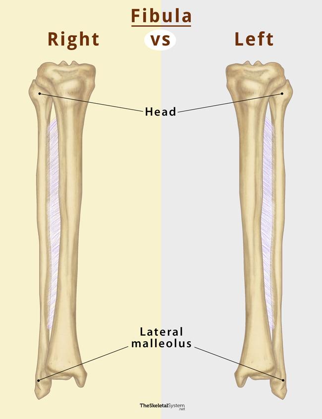
Here’s a quick way to identify the left and right fibula.
First, hold the bone so its rounded head is at the top and the pointy end is at the bottom. Now look for the bony projection and lateral malleolus on the lower end. If the lateral malleolus lies on the left, it is a right fibula and vice versa.
FAQs
Ans. No, the fibula is not a weight-bearing bone.
Ans. Yes, the fibula is a part of the knee joint.
References
- The Fibula – Teachmeanatomy.info
- Fibula – Innerbody.com
- Fibula – Radiopaedia.org
- Fibula – Kenhub.com
- Anatomy, Bony Pelvis and Lower Limb, Fibula – Ncbi.nlm.nih.gov