Calcaneus (Heel Bone)
What is the Calcaneus Bone
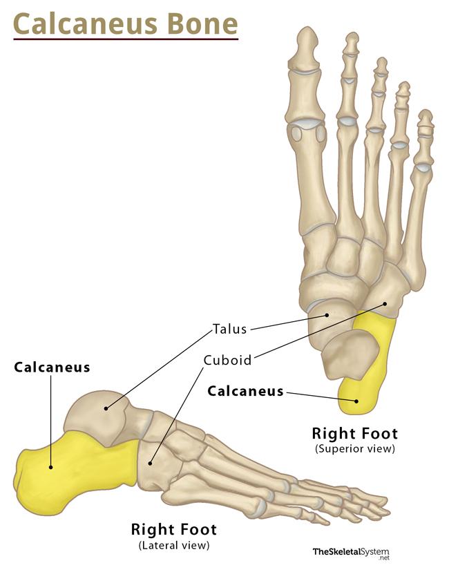
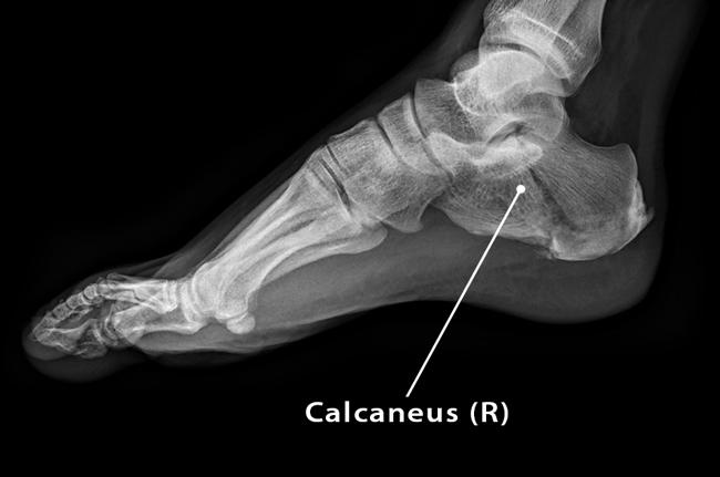
Calcaneus also called the heel bone, is an irregularly-shaped, short, cuboidal bone located in the hindfoot, just below the ankle. It is the largest tarsal, as well as the largest foot bone that forms the prominence of the heel. It is a weight-bearing bone, supporting the entire body weight while standing or walking.
The term ‘calcaneus’ is derived from the Latin word ‘calcaneum‘, meaning ‘heel’.
Where is the Calcaneus Located
Calcaneus is located in the hind region of the foot, specifically in the heel region. More specifically, it lies below the three bones, the talus, tibia, and fibula, that make up the ankle joint. Whenever you touch your heel, you can feel the calcaneus.
Calcaneus Facts
| Type | Short bone |
| Length | Approximately 75 mm |
| Number in the human body | 2 (1 in each foot) |
| Articulates with | Two other tarsal bones: talus and cuboid. |
Functions
- Bear the body weight while walking or standing.
- Transfer most of the body weight from the leg to the foot.
- Act as a lever for the calf muscles.
Anatomy – Parts and Bony Landmarks of the Calcaneus
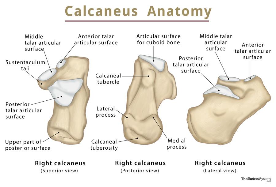
As stated, the calcaneus is an irregular cuboid bone whose superior surface can be divided into three areas – the posterior, middle, and anterior regions.
1. Posterior Region
The posterior part of the calcaneus is large, rough, convex, and dome-shaped. The Achilles or calcaneal tendon gets inserted on the superior side of the posterior part. In the front and back of this insertion point, i.e., internally and externally, there are two bursae(fluid-filled sacs), that act as cushions. The convexity of the posterior part supports the Kager’s fat pad, the fibroadipose tissue between the calcaneal tendon and the ankle joint. This part also features a thickened landmark called the calcaneal tuberosity that provides the origin site for the abductor digiti minimi and abductor hallucis. The tuberosity bears the medial and lateral processes.
2. Middle Region
The middle part bears three articular surfaces: the anterior talar articular surface, the middle talar articular surface, and, the largest, the posterior talar articular surface. The calcaneus articulates superiorly with its adjacent tarsal bone talus through these three surfaces. A rough depression, called the calcaneal sulcus or sulcus calcanei, narrows into a groove connecting the sinus tarsi with the talus on the medial side. On the lateral side of the calcaneus, a tubercle known as the peroneal or calcaneal tubercle is present.
3. Anterior Region
The front of the calcaneus, the part lying on the side of the toes, is roughly triangular. It bears a saddle-shaped articular surface that articulates with another tarsal bone, the cuboid. A horizontal shelf-like structure arises on the anteromedial portion of the bone, called the sustentaculum tali.
Development and Ossification
The calcaneus develops from two ossification centers: one primary and another secondary. The primary center starts to form during the 3rd month of the gestational period, and the other one usually after age 8. The whole process of ossification gets completed by the age of 15.
Joints and Articulations
1. Talocalcaneal or Subtalar Joint: A synovial joint, formed betweencalcaneus and talus.
2. Calcaneocuboid Joint: Another synovial joint present between calcaneus and cuboid.
Muscle and Ligament Attachments
As the calcaneus is the largest foot bone, it provides a large surface area for the complex attachment of the following muscles:
- Triceps surae (gastrocnemius and soleus) : Gets inserted in the calcaneal tubercle through the calcaneal tendon
- Abductor halluces: Originates from the medial process of the calcaneal tuberosity.
- Flexor digitorum brevis: Originates from the medial process of calcaneal tuberosity.
- Quadratus plantae: Originates from the plantar surface of the calcaneus.
- Abductor digiti minimi: Originates from the medial and lateral process of the calcaneal tuberosity.
- Extensor digitorum brevis: Originates from the dorsolateral surface of the bone.
- Extensor hallucis brevis: Originates from the sinus tarsi.
Identification of Left and Right Calcaneus
A simple way to identify the left and right calcaneus is as follows:
First, hold or place the bone in a way so that the superior convex surface is up. In this position, the smooth anterior surface would be on the outer side, and the posterior surface, i.e., the side of the heel, would be on your side.
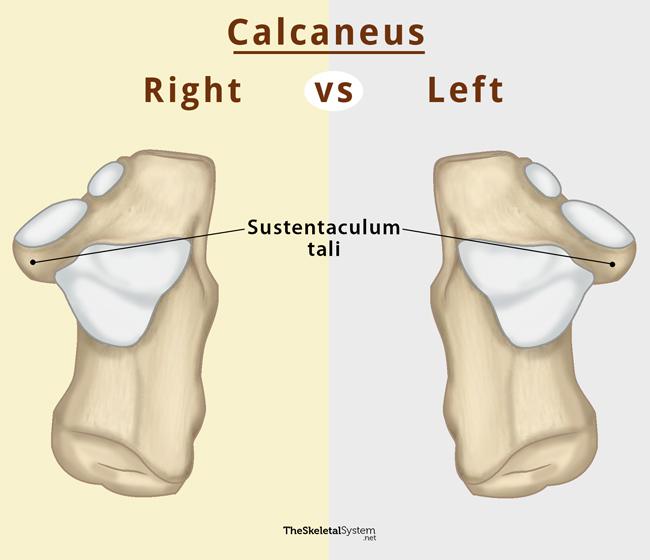
Carefully look for the bony projection on the medial side, the sustentaculum tali. If it lies to the left, it is the right calcaneus and vice versa.
References
- Calcaneus – Radiopaedia.org
- Calcaneus – Kenhub.com
- Calcaneus – Healthline.com
- Calcaneus – Sciencedirect.com