Talus
Table of Contents:
What is the Talus Bone
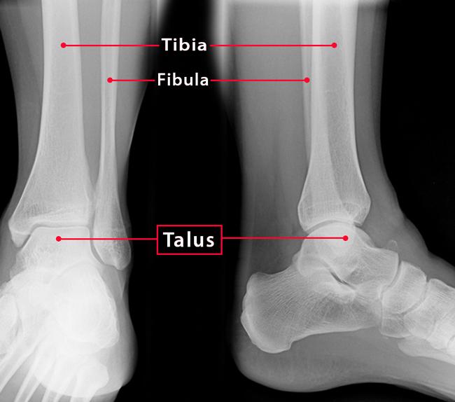
Talus bone, alternatively known as talus, ankle bone, or astragalus, is the second-largest tarsal bone that connects the leg to the foot by forming the ankle joint. This short, irregular, saddle-shaped bone gets its name from the Latin word ‘taxillus’, meaning a ‘small die or cube’, as this bone from horses was used to make dice for various games of chance by Roman soldiers.
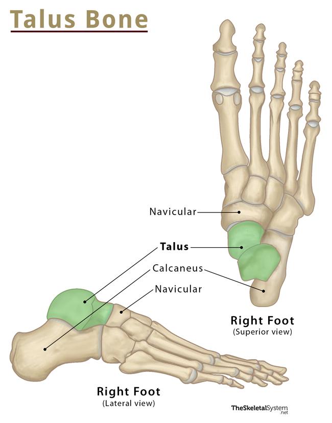
Where is the Talus Located
Talus is located in the hindfoot region, between the heel bone (calcaneus) and the tibia and fibula of the lower leg.
Quick Facts
| Type | Short bone |
|---|---|
| Length and Width | Approximately 65.15 mm Approximately 48.4 mm |
| How many are there in the human body | 2 (1 in each foot) |
| Articulates with | Four bones: the tibia, fibula, calcaneus, and navicular |
Functions
- The talus acts as the main connector between the foot and leg, forming the ankle joint.
- It allows the connecting bones of the ankle to slide around it in multiple directions while supporting the body’s weight.
- Its primary function is to transmit body weight from the tibia to the heel bone (calcaneus), thus enabling a person to maintain balance while walking.
- It takes on the extra force when the foot gets twisted, or any weight is applied to it suddenly, particularly in the ankle region.
Structure and Anatomy
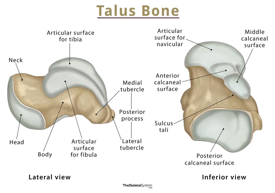
As mentioned, this is a short, irregular, saddle-shaped bone, which can be divided into three parts: head, neck, and body. The talus also bears several articular surfaces and two protuberances on the back and side, the posterior and lateral processes. It is wider in front than in the back. Its two-thirds part remains covered with cartilage to help cushion the movements it makes as part of the ankle and the foot.
Bony Landmarks
Head
The part of the talus that lies towards the toe is referred to as the talar head. It is large, oval, and carries a convex anterior surface in front for articulating with the navicular bone. On its lower side, the inferior surface bears three articular areas, separated by smooth ridges.
The head has medial and lateral facets for articulation with the calcaneum. The medial articular surface is convex, being triangular or semi-oval in shape, whereas the lateral, also called the anterior calcaneal articular surface, is a bit flattened. In between these two facets, there is another one through which the plantar calcaneonavicular ligament or spring ligament runs.
Neck
The neck of the talus is the constricted region between the oval head and body. It bears several rough surfaces for the attachment of ligaments. Its inferior surface contains a deep groove, the sulcus tali, which forms the roof of the sinus tarsi upon articulating with calcaneus. The sinus tarsi is a small tunnel located between the talus and calcaneus bones, containing numerous ligaments and a joint capsule. At the back of the sulcus tali is a large facet that articulates with the calcaneus.
The relatively thin diameter of the neck makes it a weaker area and hence more vulnerable to fractures.
Body
The cuboid body of the talus comprises most of its volume. It bears five surfaces: superior, inferior, medial, lateral, and posterior.
The superior surface also called the trochlear surface or talar dome, is curved and smooth. This part of the bone articulates with the tibia of the lower leg. It remains covered with hyaline cartilage, convex from front to back, slightly concave from side to side, and broader in front than behind.
The medial surface bears a pear-shaped articular facet for the tibial end (the medial malleolus), in continuation with the trochlear surface. Below the articular surface, there is a rough depression for the attachment of the deltoid ligament.
The lateral surface carries a large triangular concave facet for articulation with the lower end of the fibula (the lateral malleolus). Its front half is continuous with the trochlear surface and has a rough depression for the attachment of the anterior talofibular ligament. The lower part of this surface forms a bony projection called the lateral process that supports the lower portion of the lateral articular facet.
The posterior surface has a posterior process, which has a lateral and medial tubercle separated by a groove through which the tendon of flexor hallucis longus runs.
Articulations
Ankle joint or talocrural joint: It is a hinge joint, where the talus articulates with the leg bones, tibia, and fibula.
Talocalcaneonavicular joint: The following two synovial ball and socket joints are collectively known as talocalceneonavicular joint.
- Subtalar or talocalcaneal joint: Here, the talus joins the heel bone below.
- Talonavicular joint: Here, the talus joins the navicular in front.
Ligament Attachments
Though no muscles are attached to the talus, many ligaments are attached to this bone. These ligaments ensure that the talus cannot rock from side to side or move backward or forward relative to the tibia and fibula.
On the sides, the ankle joint is held together by the posterior talofibular and anterior talofibular ligaments. From the center, it is held together by a massive ligament, the deltoid ligament.
All the ligaments attached to talus are listed below:
- Anterior talofibular ligament
- Posterior talofibular ligament
- Talocalcaneal ligaments
- Tarsal sinus ligaments
- Cervical ligament
- Talocalcaneal interosseous ligament
- Deltoid ligament
- Anterior tibiotalar ligament
- Posterior superficial tibiotalar ligament
- Posterior deep tibiotalar ligament
- Dorsal talonavicular ligament
FAQs
Ans. No, the talus is not a weight-bearing bone. It transmits the entire body weight to the foot.
References
- Talus – Kenhub.com
- Talus – Radiopaedia.org
- Anatomy of the Talus Bone – Study.com
- Talus – Sciencedirect.com
- Anatomy, Bony Pelvis and Lower Limb, Foot Talus – Ncbi.nlm.nih.gov
- Bones of the Foot: Tarsals, Metatarsals and Phalanges – Teachmeanatomy.info