Tibia (Shin Bone)
What is the Tibia
The tibia, also known as the shank or shin bone, is one of the two long leg bones of the lower leg. It is a weight-bearing bone.
Where is the Tibia Located
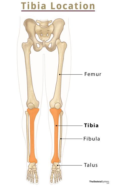
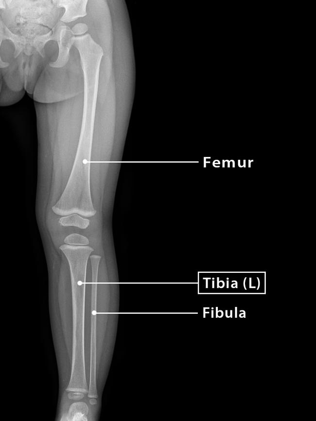
As mentioned, the tibia is located in the lower leg, extending from the knee to the ankle. More precisely, it is situated on the distal side of the femur and the proximal side of the talus of the foot. The tibia is also located medially to the other bone of the lower leg, called the fibula.
You can feel the presence of this bone by touching the front portion of your lower leg, just below the knee.
Quick Facts
| Type | Long bone |
| Length | Approximately 36 cm |
| How many are there in the human body | 2 (1 in each leg) |
| Articulates with | Femur, fibula and talus |
Functions
- Support body weight while standing or during any activity.
- Act as a lever for the leg during movements like walking, running, jumping.
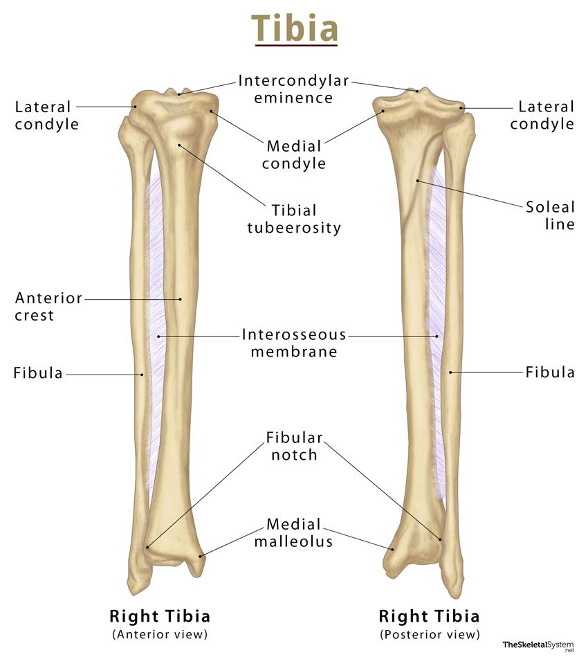
Anatomy – Parts of the Tibia
It is a long bone with two ends, proximal and distal, and an intervening shaft. The part lying on the side of the knee is known as the proximal tibia, whereas the part lying on the side of the foot is known as the distal tibia.
Proximal Tibia and its Bony Landmarks
As mentioned, the proximal tibia refers to the upper end of the bone. It is roughly flat and possesses some important bony landmarks.
1. Medial and Lateral Condyles: The proximal tibia bears two prominent condyles. The one facing the midline of the body, or medially, is referred to as the medial condyle. On the other hand, the one facing towards the outer side of the body, or laterally, is called lateral condyles. They articulate with the femoral condyles, forming the knee joint. Out of the two condyles, the medial is larger than the lateral.
2. Tibial plateau: The top surface of the two condyles form a flat surface, known as the tibial plateau.
3. Intercondyloid eminence: In between the two condyles is a region called the intercondylar eminence or tibial spine. It projects upwards on either side as the medial and lateral intercondylar tubercles. The lateral tubercle is commonly known as the Gerdy tubercle.
4. Intercondylar area: As the name suggests, it is the region between the two condyles.
The Shaft and its Bony Landmarks
The shaft or body is the region between the two extremities of the tibia. It appears triangular in cross-section, with three borders: anterior, medial, lateral, or interosseous. These three borders form three surfaces; the medial, lateral, and posterior.
Borders
1. Anterior border: It is the most prominent border, which can be observed as a distinct margin that continues distally from the tibial tuberosity. It is also known as the anterior crest, which begins above the tibial tuberosity, and ends below at the anterior margin of the medial malleolus. The anterior border is the part of the tibia that can be felt by touching the front portion of the lower leg.
2. Medial border: The medial border commences at the posterior region of the medial condyle and extends up to the posterior border of the medial malleolus.
3. Lateral border or interosseous crest: It begins below the iliotibial tract’s tubercle and descends the tibia’s lateral surface. The lateral border gives attachment to the interosseous membrane that binds the tibia and the fibula together. The fibular notch replaces the interosseous border at the lower end of the tibia. The distal end of the fibula fits here.
Surfaces
1. Medial surface: The medial surface is smooth, convex, and broader than below. It remains bound by the anterior and medial borders. It is subcutaneous, meaning there is a minimal fat layer between the bone and the skin and has no muscles attached to it. Due to this, the medial surface is palpable along the entire length of the leg, specifically on the anteromedial side.
2. Lateral surface: The lateral surface is narrower than the medial. The anterior and interosseous margins border it.
3. Posterior surface: The posterior surface is confined by the interosseous and medial margins. It features a bony ridge called the soleal line. The line crosses this surface diagonally and eventually blends with the medial border of the tibia.
Distal Tibia and its Bony Landmarks
At the distal end, the tibia widens and appears rectangular in cross-section. It has two bony landmarks, the medial, malleolus, and fibular notch.
1. Medial malleolus: The medial surface of the distal end features a bony projection called the medial malleolus. It articulates with the talus to form part of the ankle joint.
2. Fibular notch: The lateral surface of the distal end features a facet for the distal end of the fibula, called the fibular notch. The tibia and fibula join to form the distal tibiofibular joint at this spot by a thickening of the interosseous membrane.
Articulations
1. Superior tibiofibular joint: Here, the proximal end of the tibia articulates with the head of the fibula, forming a synovial joint.
2. Middle tibiofibular joint: It is a fibrous joint formed by the interosseus membrane, which spans between the shafts of the tibia and its adjacent bone, fibula, attaching to the interosseous margins of each bone.
3. Inferior tibiofibular joint: It is formed by connecting the fibular notch of the distal end of the tibia with the distal end of the fibula.
All these three joints hold the tibia and fibula together.
Muscles Attached
Tibia has many muscles attached to it. Some originate from the bone, whereas some get inserted into it.
Originating from the Tibia
1. Tibialis anterior – Lateral surface of the tibia
2. Extensor digitorum longus – Lateral condyle of the tibia
3. Tibialis posterior – Posterior surface of the tibia
4. Soleus – Posterior surface of the tibia on the soleal line
5. Flexor digitorum longus – Posterior surface of tibia on the soleal line
Inserting on the scapula
1. Sartorius and gracilis – Medial surface of proximal tibia
2. Quadriceps femoris – Tibial tuberosity
3. Semimembranosus – Medial condyle of tibia
4. Semitendinosus – Proximal end of the tibia below medial condyle
5. Popliteus – Soleal line of the posterior tibia
6. Tensor fasciae latae – Lateral tubercle of the tibia
Identifying the Left and Right Tibia Bones
Here’s a quick way to differentiate between the left and right tibia.
First, hold the bone vertically so that the medial and lateral condyles are at the top and the bony projection, the medial malleolus, is at the bottom.
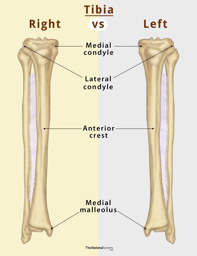
Now, identify the larger medial condyle and see which side it faces. If it faces your left, it is a right tibia and vice versa.
Alternatively, you can also observe which side the medial malleolus lies on. If it lies on the right side, it is a right tibia and vice versa.
FAQs
Ans. Talus articulates with the tibia.
References
- The Tibia – Teachmeanatomy.info
- Tibia – Kenhub.com
- Tibia – Innerbody.com
- Tibia – Radiopaedia.org
- Anatomy, Bony Pelvis and Lower Limb, Tibia – Ncbi.nlm.nih.gov