Navicular Bone
What is the Navicular Bone
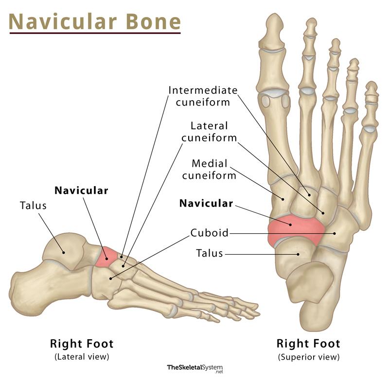
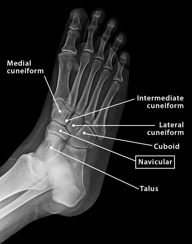
Navicular is one of the seven tarsal bones located in the midfoot region. In Latin, ‘navicular’ means ‘a little ship’. So, the bone got its name from Latin due to its resemblance to a small boat. It connects the ankle bone or talus to the other two tarsal members, the cuboid and the three cuneiforms.
Where is it Located
Navicular bone is located on the medial side of the midfoot, anterior to the talus, posterior to the cuneiform bones, and next to the cuboid bone.
Navicular Bone Facts
| Type | Short bone |
| Length | Approximately 3.5 cm |
| Number in the human body | 2 (1 in each foot) |
| Articulates with | Talus, cuboid, and three cuneiforms |
Functions
- Stabilize the ankle and maintain the arch of the foot while walking.
Structure and Anatomy
Navicular is a short, irregular, boat-shaped bone. It bears some articular surfaces for the attachment of adjacent tarsal bones and a few other bony landmarks.
Bony Landmarks
Anterior Surface
The anterior surface of the navicular bone is convex and kidney-shaped, which divides into three articular surfaces by two faint ridges. The articular surfaces are for the attachment of three cuneiform bones, medial, intermediate, and lateral.
- The medial articular surface is the largest of the three articular surfaces. It is convex and has a triangular-shaped surface, articulating with the medial cuneiform bone.
- Unlike the medial surface, the intermediate articular surface is flat but has a triangular shape. It articulates with the intermediate or middle cuneiform bone.
- The lateral articular surface is the smallest among the three. It has a quadrangular surface and articulates to the lateral cuneiform bone.
These three articulations converge at the plantar aspect and form the arch of the foot.
Posterior Surface
The posterior surface of the navicular bone is concave and remains covered with articular cartilage that connects with the head of the talus.
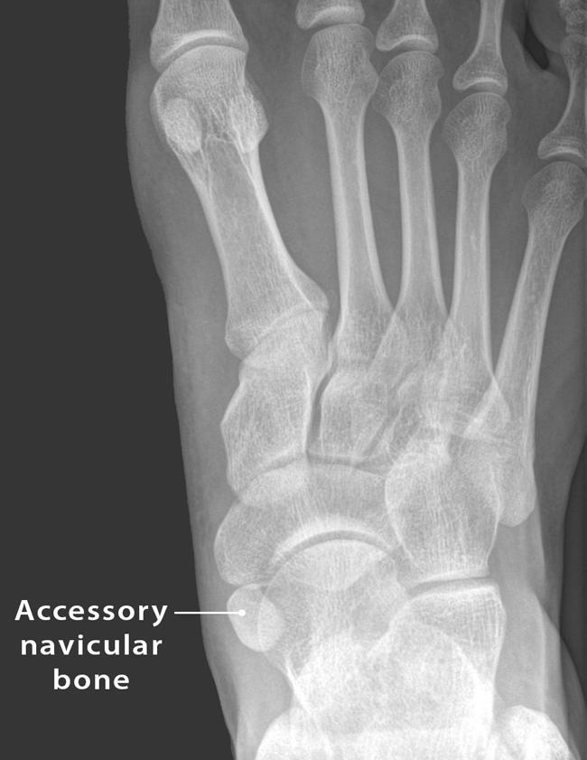
Medial Surface
On the medial side of the bone, there is a bony projection called navicular tuberosity. An accessory or supernumerary bone may sometimes exist adjacent to the navicular tuberosity, giving a bony prominence on the medial side. This bone is called the accessory navicular bone. It is a congenital situation, present from birth.
Lateral Surface
The lateral surface is irregular, containing a facet for articulation with the cuboid.
Joints and Articulations
1. Talonavicular joint: This joint is formed between the talus and navicular bone.
2. Cuneonavicular joint: The navicular bone articulates with the three cuneiform bones, medial, intermediate, and lateral, to form this joint.
3. Cuboideonavicular joint: This joint is formed between the navicular and cuboid bones.
Muscle and Ligament Attachments
The only muscle attached to the navicular is the tendon of the tibialis posterior. It gets inserted into the bone medially on the navicular tuberosity.
Several ligaments attach to this bone.
- The talonavicular ligament attaches the dorsal surface of the bone to the neck of the talus.
- The plantar calcaneonavicular or spring ligament is a group of ligaments that bind the navicular and the calcaneus to form a socket for the head of the talus.
- The dorsal cuneonavicular ligament and plantar cuneonavicular ligament join each cuneiform bone to the navicular.
References
- Navicular bone – Kenhub.com
- Anatomy, Bony Pelvis and Lower Limb, Navicular Bone – Ncbi.nlm.nih.gov
- Bones of the Foot: Tarsals, Metatarsals and Phalanges – Teachmeanatomy.info
- Navicular – Radiopaedia.org
- Navicular Bone – Sciencedirect.com