Radius
Published on September 7th 2018 by Danielle Budd
Definition: What is the Radius
The radius, also known as the radial bone, is one of the two forearm bones in the human body, with the other being the ulna. It is instrumental in the shaping and use of hands [1].
Where is the Radius Bone Located in the Arm
It is located on the thumb side of the hand, lying laterally in the lower arm, parallel in reference to the ulna [1, 2].
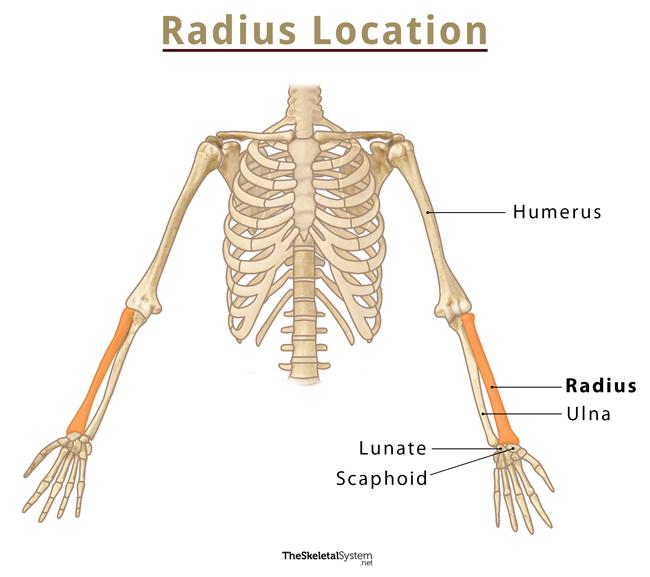
Radius Bone Location
Radial Bone Facts |
|
| Number in the human body | Two ― one in each arm |
| Type | Long bone [2] |
| Primary ossification centers | One ― at the middle of the shaft (appearing around the 8th week of fetal life) [3] |
| Secondary ossification centers | Two ― one for the distal end (appearing around 2 years) and another for the proximal end (appearing around 5 years) (all the centers fuse together by 20 years) [4] |
| Forms articulations with | Humerus, ulna, scaphoid, lunate |
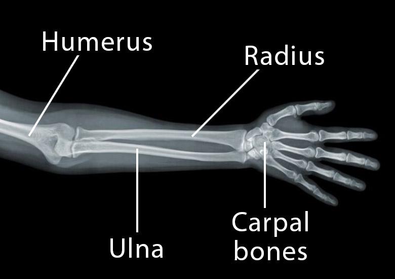
Radius X-ray Picture
Functions of the Radius
Proper functioning of the radius is essential for performing any day-to-day activity with our hand, from holding something, balancing with the arm, throwing something, writing, typing, using the phone, etc.
- It forms a hinge joint with the humerus bone, which allows us to flex and extend the elbow [7].
- The radius moves around the ulna at the wrist, enabling us to turn our hand’s palm up and down [8].
- The bone also forms an ellipsoidal joint with the proximal carpal row that allows us to move, rotate, bend, and flex the wrist [7].
Radius Bone Anatomy
The Radial bone is somewhat triangular longitudinally [2], divided into the upper end, body/shaft, and the lower end.
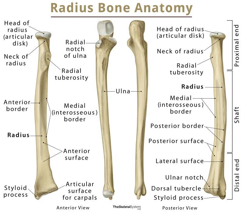
Radius Bone Anatomy Labeled Diagram
Parts of the Radius |
|
1. Upper End (Proximal Radius) |
|
| Landmarks:
1. A disk-shaped head (caput radii) 2. A neck, continuing from the head, narrowing towards the shaft [2] 3. The radial tuberosity, a bony projection below the neck [3] |
Surfaces and Articulations:
1. A concave articular surface on top of the head for the capitulum of the humerus (elbow humeroradial joint) [5] 2. A smooth circumference of the head articulating with the radial notch of the ulna (proximal radio-ulnar joint) [3] |
2. Body/Shaft |
|
| Landmarks:
There are several landmarks on the radial shaft for the origin and insertion of various tendons [6] |
Borders
1. Anterior; 2. Posterior; 3. Medial (or interosseous, the sharpest border where the interosseous membrane connects) Surfaces 1. Anterior; 2. Posterior; 3. Lateral [3] |
3. Lower End (Distal Radius) |
|
| Landmarks:
1.A styloid process projecting distally on the lateral side 2. A prominent dorsal tubercle (or Lister’s tubercle) on the dorsal surface [3] 3. The ulnar notch on the medial side [1] |
Surfaces and Articulations:
1. The concave surface of the ulnar notch articulating with the ulnar head (distal radio-ulnar joint) [2] 2. A lateral triangular area on the distal or inferior surface forming joints with the carpal bones, scaphoid, and lunate (wrist joint) [2] |
Radius Bone Muscle Attachments |
|
Name of Muscle |
Insertion at Radius |
| Biceps brachii | The rough posterior surface of the radial tuberosity [3] |
| Pronator teres | Lateral surface of the shaft |
| Pronator quadratus | Medial surface of the shaft |
| Supinator | Laterally on the shaft, covering one-third of the proximal radius (both origin and insertion) |
Name of Muscle |
Origin at Radius |
| Flexor digitorum superficialis | Medial surface of the shaft |
| Flexor pollicis longus | Medial surface of the shaft |
| Abductor pollicis longus | Anterior surface of the shaft [5] |
There is a layer of hyaline cartilage covering both the proximal and distal ends of the radius. This makes the articular surfaces smoother so there is less friction in the joints during arm movements. It also works as a shock absorbent to reduce stress on the elbow and wrist joints from any impact [1].
The anterior part of the radial tuberosity is covered in a synovial bursa, called the radial bursa, to keep it separated from the biceps tendons (of the biceps brachii muscle) during movements [3].
Radius Bone Side Determination
When the radial tuberosity is facing anteriorly (or facing you), the styloid process of the radius should be on the same side as the thumb. Holding the bone in this manner helps determine whether it is the left or right radius.
FAQ
What are the most common injuries and conditions associated with the radius?
The radius is considered the most commonly fractured bone in the human body, with distal radius fractures being the most common form of radial fracture [9]. Radial head dislocation is another common injury associated with the bone [10]. The bone may also be affected by arthritis of the wrist or elbow joints.
Ulna or radius ― which bone is longer and larger?
Though the ulna is longer than the radius, the latter is comparatively thicker throughout its length, especially in the shaft area [8].
References
- http://www.innerbody.com/image_skel14/skel20.html
- http://teachmeanatomy.info/upper-limb/bones/radius/
- https://www.earthslab.com/anatomy/radius-bone/
- https://www.bartleby.com/107/53.html
- https://www.kenhub.com/en/library/anatomy/the-radius-and-the-ulna
- https://courses.lumenlearning.com/boundless-ap/chapter/the-upper-limb/
- https://www.getbodysmart.com/upper-limb-bones/radius-ulna
- https://radiopaedia.org/articles/radius
- https://orthoinfo.aaos.org/en/diseases–conditions/distal-radius-fractures-broken-wrist/
- https://www.healthline.com/human-body-maps/radius-bone