Humerus
What is the Humerus Bone
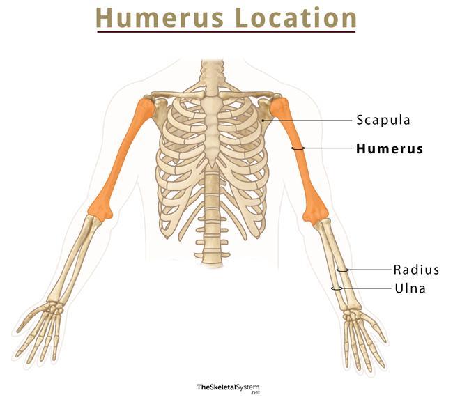
The humerus is a long bone in the human arm, running from the shoulder to the elbow. It is the largest bone in the human arm, and the only one in the upper arm, sometimes referred to as the upper arm bone. The humerus acts as the attachment point for multiple powerful muscles and helps with all arm activities, like writing, lifting, and throwing.
Being one of the longest bones in the body, it is more prone to fractures upon impact.
Where is the Humerus Located
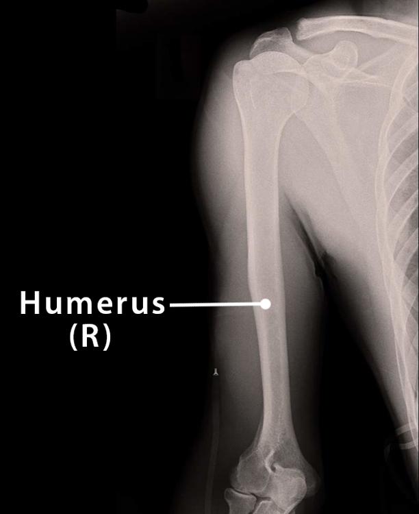
The humerus is located between the shoulder and elbow joints in the upper arm.
Humerus Facts
| Type | Long bone |
| Length | 14.4 inches (on average in adults) |
| How many are there in the human body | 2 (1 in each arm) |
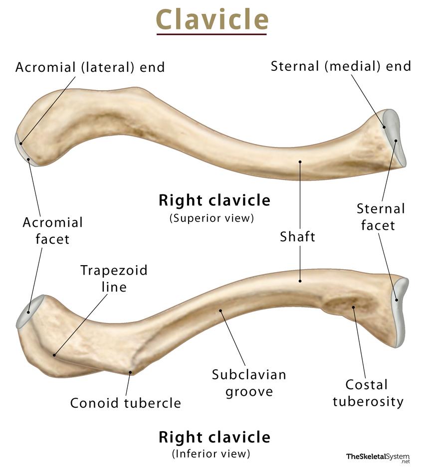
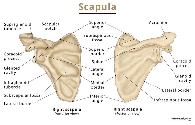
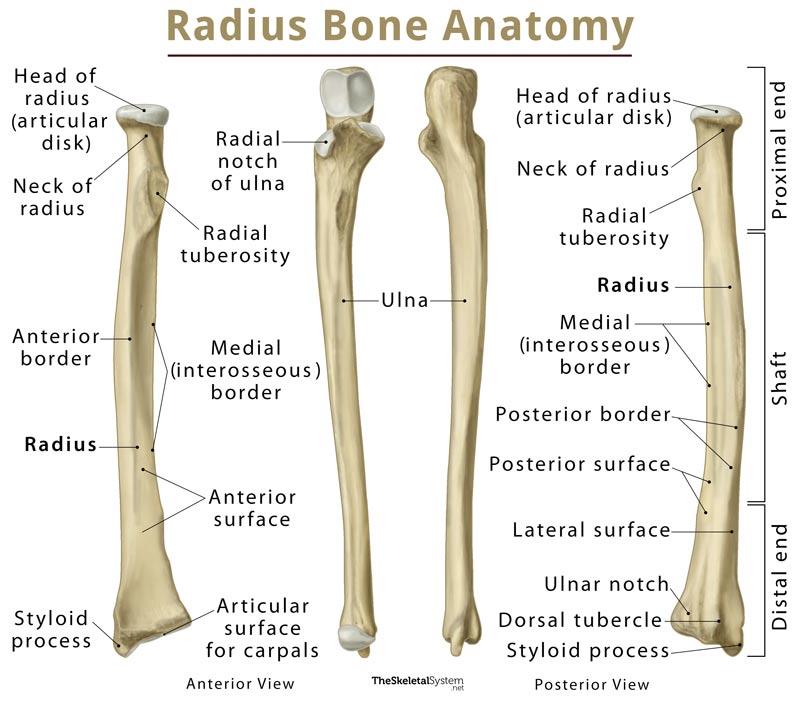
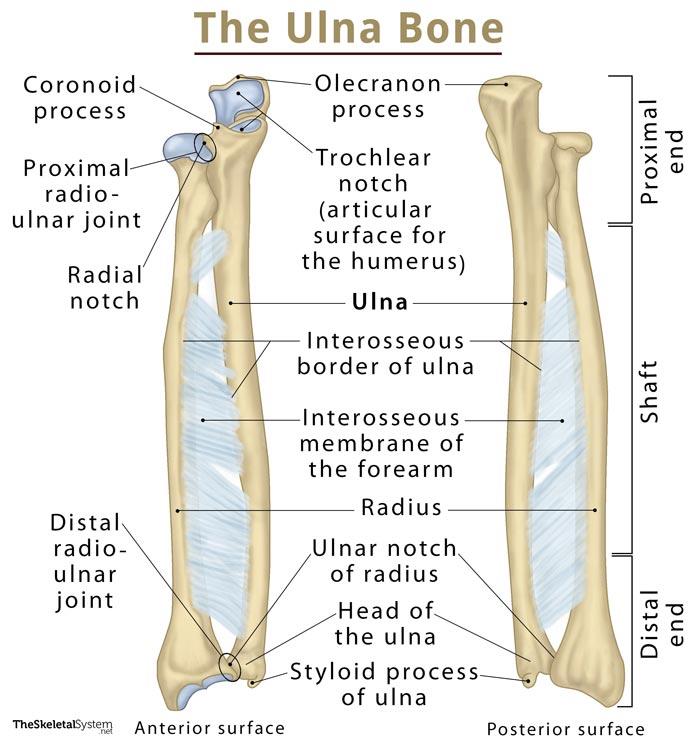
| Articulates with | Shoulder blade or scapula (proximal side), Ulna and radius (distal side) |
Functions
- It helps in the functioning of the upper limb by providing structural support, and serving as an attachment site to 13 muscles that aid in the movements of the hand and elbow.
- The humeral head makes up a portion of the ball-and-socket shoulder joint, which is the insertion point for muscles making up the shoulder girdle.
- Several ligaments present in this area assist with securing the musculature. They also provide motion to the shoulder joint.
- The basilic vein, traveling close to the humerus, helps drain parts of the hand and forearm.
- The brachial plexus lying across the front portion of the bone provides sensation and motion to every muscle in the arm and certain portions of the neck and spinal cord.
Structure and Anatomy
The humerus comprises a proximal region, a shaft, and a distal region. All of these are important anatomical landmarks.
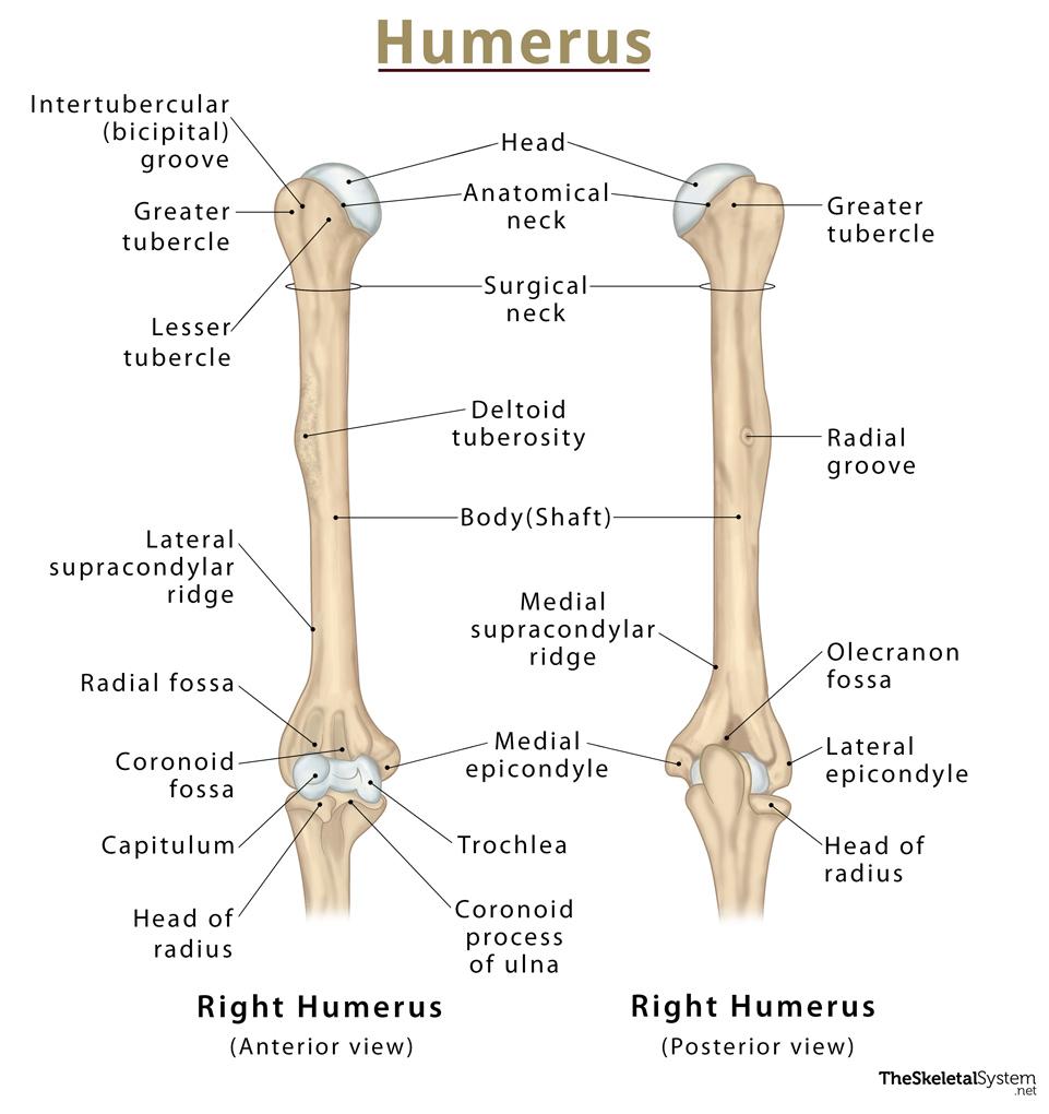
The detailed anatomy of each part is discussed below:
1. Proximal Landmarks of Humerus
Head: The proximal end of the humerus forms a smooth, spherical structure known as the head. It is the ball in the the ball-and-socket joint in the shoulder, where the glenoid cavity of the scapula acts as the socket. Due to its round shape, the head rotates around its axis at the shoulder joint and moves in all directions.
Anatomical Neck: Just below the head, the humerus narrows into the anatomical neck. It separates the head from the other two regions: greater and lesser tuberosities.
Greater Tubercle (Greater Tuberosity): It lies on the lateral side of the bone, with an anterior, and a posterior surface. The three rotator cuff muscles, supraspinatus, infraspinatus, and teres minor, attach to the greater tubercle’s superior, middle, and inferior facets, respectively.
Lesser Tubercle (Lesser Tuberosity): It is much smaller than the greater tubercle located inferior to the head, bearing only an anterior surface. This is where the the fourth and last rotator cuff muscle, the subscapularis, attaches.
Intertubercular Sulcus: Separating the two tuberosities is a deep groove known as the intertubercular sulcus or bicipital groove. The tendon of the bicep’s long head emerges from the shoulder joint and runs through this groove. The edges of the intertubercular sulcus are known as lips, where pectoralis major, teres major, and latissimus dorsi get inserted.
Surgical Neck: It is the part between the tuberosities and the shaft where the humerus narrows down to form the surgical neck before extending toward the elbow joint. Here, circumflex humeral vessels and the axillary nerve lie against the bone.
2. The Shaft
A long, cylindrical shaft (body) composes the middle part of the humerus. It bears a roughened surface on the lateral side, known as the deltoid tuberosity, as the deltoid muscle gets attached there. The width of the bone gradually increases past the deltoid tuberosity, becoming double as it moves towards the elbow joint.
A shallow depression called the radial (or spiral) groove runs diagonally down the posterior surface of the humerus, parallel to the deltoid tuberosity. This groove contains the radial nerve and the profunda brachii artery.
The shaft serves as the surface for attachment of several muscles
Anterior Side: Coracobrachialis, brachialis, deltoid, brachioradialis
Posterior Side: The medial and lateral heads of the triceps. Their origin is marked by the spiral groove on the posterior side of the humerus.
3. Distal Region of Humerus
The lower end of the humerus is the distal humerus, containing two joint-forming processes, the capitulum and the trochlea. The trochlea tightly hinges with the forearm’s ulna, forming half of the elbow joint. On the other hand, the convex capitulum articulates with the concave radial head on the lateral side of the arm. The joint thus formed is the elbow that allows the human arm to bend and fold in the middle.
A small cavity called the olecranon fossa on the posterior side of the bone locks the olecranon or the tip of the ulna into the bone. This locking prevents us from extending the elbow beyond 180 degrees. Despite this, the distal portion of the humerus also contains two other depressions, known as the coronoid and radial fossae. They lodge the forearm bones during flexion or extension of the elbow.
The medial and lateral borders of the distal humerus form the medial and lateral supracondylar ridges. Among the two ridges, the lateral supracondylar ridge is relatively roughened, providing the common origin site of the forearm extensor muscles.
The extracapsular projections of the bone, the lateral and medial epicondyles, are found immediately distal to the supracondylar ridges. Both of the epicondyles can be felt at the elbow from the outside. Among the two, the medial is larger, extending more towards the elbow joint. The ulnar nerve passes through a groove present on the posterior side of the medial epicondyle.
Articulations
Proximally: The shoulder (or glenohumeral) joint is formed where the humerus articulates with the scapula’s glenoid cavity.
Distally: The elbow joint is formed where it articulates with the trochlear notch of the ulna, and its capitulum articulates with the radial head.
Left vs. Right Humerus
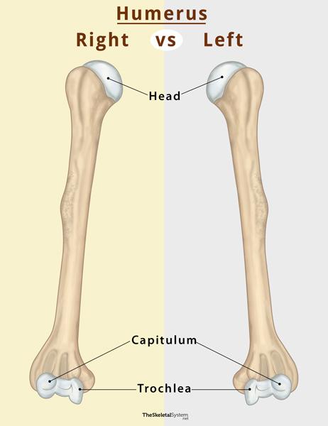
Here is how you can identify if a specific humerus bone comes from the left or right arm:
First, orient the bones in a way so that the rounded head is up. Then, look for the deep olecranon fossa on the lower (distal) end. Observe which side the capitulum is facing.
For that, first, rotate the bone so that both the capitulum and trochlea are facing you. Then, if the capitulum lies on the left side of the bone, it is a left humerus. On the contrary, if the capitulum is on the right side, it is a right humerus.
Development and Ossification
The humerus is one of the structures that begin to ossify during embryonic development. Its ossification starts from the shaft.
The humerus develops from a total of 8 ossification centers. The body starts to ossify during the 8th week of fetal development. The rest of the ossification happens after birth, starting with the head and capitulum at 1 year, the greater tubercle at 3 years, the lesser tubercle and medial epicondyle at 5 years, and the trochlea and lateral epicondyle at 10 years.
FAQs
Ans. The muscles that move the humerus include both axial and scapular muscles.
Axial muscles: pectoralis major, and latissimus dorsi
Scapular muscles: coracobrachialis, deltoid, subscapularis, supraspinatus, infraspinatus, teres minor, and teres major
Ans. When the ulnar nerve gets bumped against the humerus, one feels a strange (funny) sensation. For this reason, the humerus is called the funny bone, though it is actually the ulnar nerve that is responsible.
Ans. Being one of the longest bones in the body, it is more prone to fractures upon impact. At the proximal end, most fractures occur at the surgical neck and are common in the elderly, especially those with osteoporosis.
Other medical issues associated with the humerus include metastatic bone disease, radial nerve injury, osteochondroses, charcot arthropathy, and humerus varus.
References
- Anatomy, Shoulder and Upper Limb, Humerus – Ncbi.nlm.nih.gov
- Humerus – Sciencedirect.com
- Humerus – Radiopaedia.org
- Humerus – Osmosis.org
- The Humerus – Teachmeanatomy.info
One Response to "Humerus"