Clavicle (Collarbone)
What is the Clavicle Bone
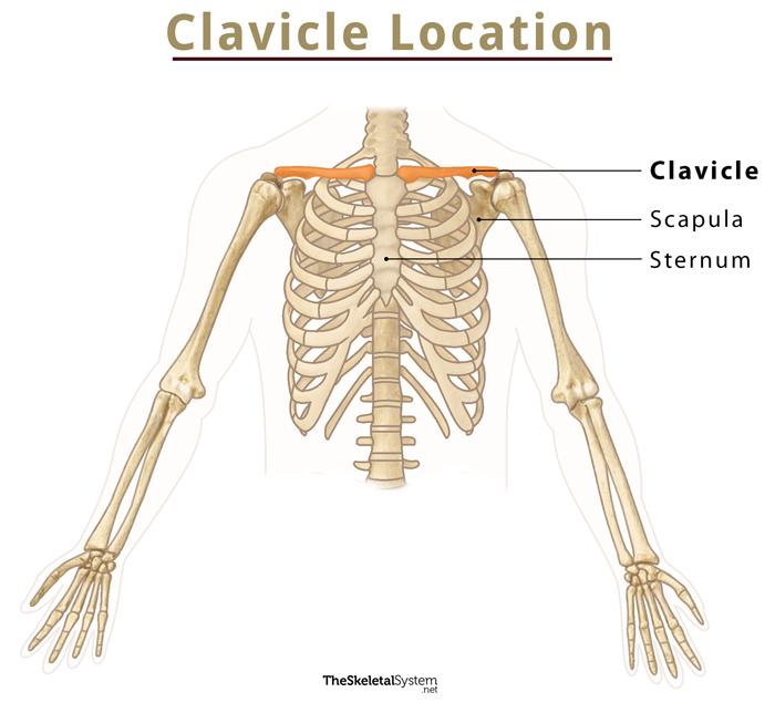
The clavicle, commonly known as the collarbone, is a slender, S-shaped, modified long bone located at the base of the neck. It is the only long bone of the body that lies horizontally.
The term clavicle comes from the Latin word ‘clavicula’, meaning ‘little key’, as its shape resembles an old-fashioned key. Also, the bone rotates along its axis like a key when the arms are moved away from the body. You can feel the presence of this bone by touching the region below your neck.
Where is the Clavicle Located
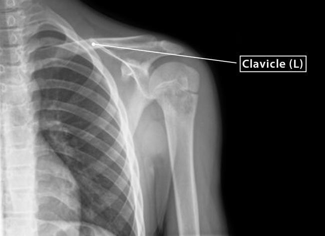
As stated, the clavicle is located at the neck’s base and across the rib cage’s upper part. It sits between the shoulder blade (scapula) and breastbone (sternum), connecting the pectoral or shoulder girdle to the axial skeleton. The clavicle is the only bone that connects the axial skeleton to the appendicular skeleton.
Clavicle Facts
| Type | Long bone |
| Length | Approximately 6 inches (15 cm) |
| Number in the human body | 2 ( 1 on either side) |
| Articulates with | Scapula and sternum |
Functions
- Connect the arm with the rest of the skeleton.
- Allow free movement of the shoulder away from the body.
- Transmit force or any physical impact from the upper limb to the axial skeleton.
- Protect the neurovascular bundle supplying the upper limb.
- Along with the rib cage, they protect the heart from external shock.
Anatomy of the Parts of Clavicle With its Bony Landmarks
Being a long bone, it has two ends, the sternal and acromial ends. The region in between the two ends is known as the shaft.
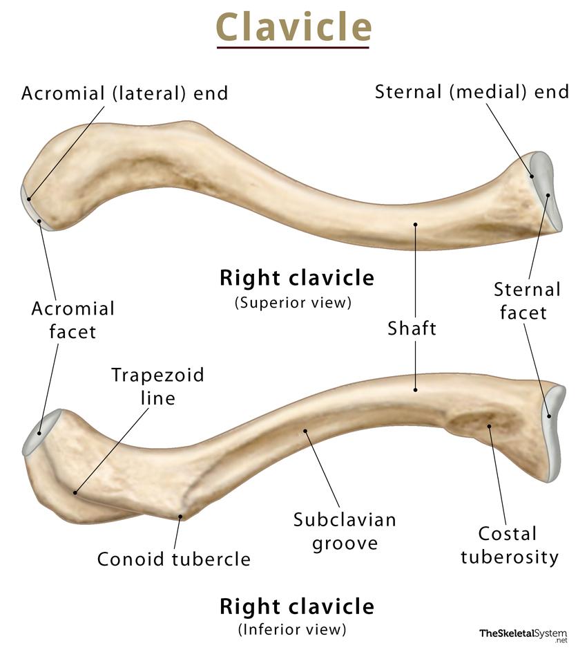
1. Sternal (Medial) End
The part of the clavicle that lies towards the sternum is called the sternal end or the medial end. It is curved and convex, bearing a rounded end that articulates with the sternum, forming the sternoclavicular joint.
i) Sternal Facet
On the far edge of the sternal end, a triangular facet, called the sternal facet is present. It bears a posterior tip and an anterior base, connecting the clavicle to the manubrium of the sternum at the sternoclavicular (SC) joint. The articular surface extends to the inferior side, articulating with the costal cartilage of the first rib.
2. Acromial (Lateral) End
The broad, flat region of the clavicle lying towards the scapula is known as the acromial end or lateral end. It is both the widest and thinnest portion of the clavicle. This region bears a facet known as acromial facet that articulates with the scapula. The lateral end has two borders: anterior and posterior. The anterior border is concave forward, whereas the posterior border is convex backward.
i) Acromial Facet
At the extreme edge of the acromial end, there is a small, flattened, and oval facet called the acromial facet. This helps the clavicle to articulate with the acromion of the scapula, forming the acromioclavicular (AC) joint.
3. Shaft
As mentioned, the middle region of the clavicle, i.e., the region between the sternal and acromial end, is the shaft. Several muscles originate from or get attached in this part.
The shaft can be divided into two regions, depending on its location and spread. The region lying on the side of the sternal end, covers the maximum portion, approximately two-thirds of the bone. This region is known as the medial two-third. On the other hand, the region of the shaft that lies towards the acromial end covers the remaining one-third of the bone. This region is known as the lateral third.
Several important bony landmarks are found in these regions.
Medial two-third
i) Costal Tuberosity
On the inferior side of the sternal end, the bone bears a roughened oval elevation, called the costal tuberosity. It is over 2 cm in length and acts as a site of attachment for costoclavicular ligament. This ligament attaches the clavicle to the costal cartilage of the first rib.
Lateral third
i) Conoid Tubercle
On the inferior surface of the bone, there is a rough, bumpy projection called the conoid tubercle. It serves as an attachment site for the conoid ligament, a part of the coracoclavicular ligament. The coracoclavicular ligament attaches the clavicle to the coracoid process of the scapula. The prominence of the conoid tubercle serves as a useful landmark to identify the inferior surface of the clavicle.
ii) Trapezoid Line
It is an elevation or ridge that runs obliquely from the conoid tubercle to the lateral end of the clavicle. It provides an attachment point for the trapezoid ligament, which is also a part of the coracoclavicular ligament mentioned above.
Subclavian groove or sulcus
It is an indentation that runs horizontally along the inferior surface of the shaft, from the costal tuberosity to the conoid tubercle. It acts as a site of attachment for the subclavius muscle.
Articulations
Sternoclavicular Joint: It is a synovial joint that is formed between the sternal end of the clavicle and the manubrium of the sternum.
Acromioclavicular Joint: It is a synovial joint that is formed between the acromial end of the clavicle and the acromion of the scapula.
Muscle Attachments
There is a total of six muscles that are attached to the clavicle. Out of these six, four muscles are attached to the sternal end or medial two-third of the clavicle, whereas two muscles are attached to the acromial end or lateral third of the clavicle.
- Sternocleidomastoid muscle: gets attached to the superior surface of the bone on the sternal end.
- Pectoralis major muscle: gets attached to the anterior surface of the bone on the sternal end.
- Subclavius muscle: present in the subclavian groove of the shaft’s inferior surface. From there, the muscle extends into both sides, lateral and medial.
- Sternohyoid muscle: attaches to the sternal end of clavicle.
- Trapezius muscle: gets attached along the posterior surface of the bone on the acromial end.
- Deltoid muscle: remains attached along the anterior surface of the bone on the acromial end.
Left and Right Clavicle – How to Identify
Here’s a quick way to distinguish between the left and right clavicle.
First, position the bone horizontally, holding its mid-region. Also, ensure that the bone is held superiorly, i.e., the smooth surface should face upwards.
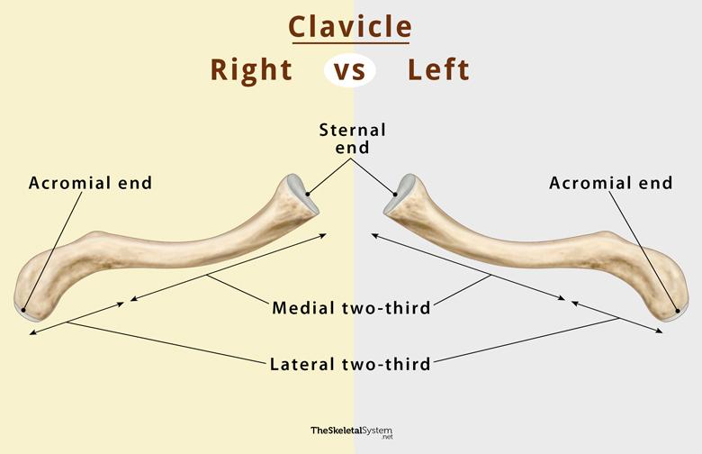
If you observe the medial two-thirds on your right side, it’s a right clavicle. On the other hand, if the medial two-thirds is on your left side, it’s a left clavicle.
FAQs
Ans. No, the clavicle is not a part of the axial skeleton.
Ans. Yes, the clavicle is a part of the appendicular skeleton.
References
- The Clavicle – Teachmeanatomy.info
- Clavicle – Kenhub.com
- Clavicle – Radiopaedia.org
- Clavicle Bone Anatomy – Getbodysmart.com
- Clavicle – Sciencedirect.com