Ulna
Definition: What is the Ulna
Ulna (plural: ulnae; pronunciation: úl-nu) is one of the two primary bones forming the forearms in humans, the other one being the radius. There is one ulna bone in each arm. It is a long bone [1] and is vital in forming both the wrist and elbow joints [2].
Where is the Ulna Bone Located in the Human Body
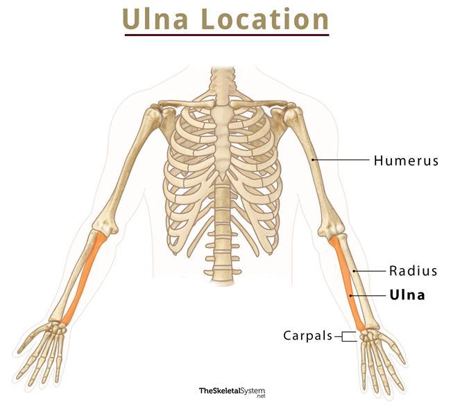
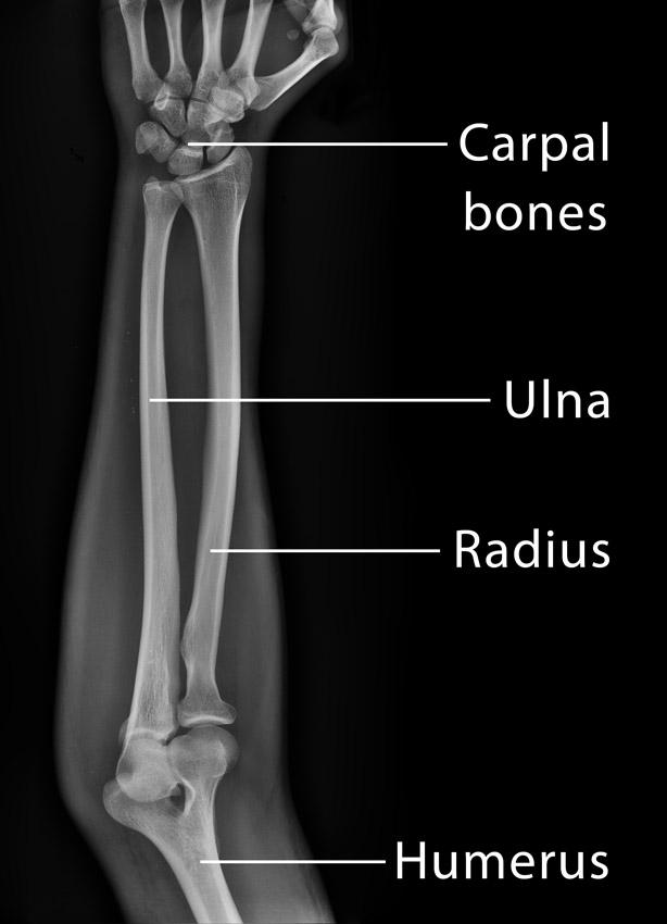
It is the medial bone of the forearm, located on the side opposite to the thumb, that is on the side of the little finger, extending from the region of the wrist to the elbow. In other words, the ulna can be found between the proximal carpal row and the upper arm bone humerus, running parallel to the other lower arm bone radius [3, 5].
Development and Ossification
The ulna ossifies from three different centers, with the primary center for the shaft appearing around the eighth week of fetal life. The secondary centers for the distal and proximal ends appear around 5-7 years and 8-10 years of age, respectively. All the centers unite when a person is around 18-20 years old [6, 7].
Anatomy of the Ulna Bone
Different Parts of Ulna and the Joints Formed by Them
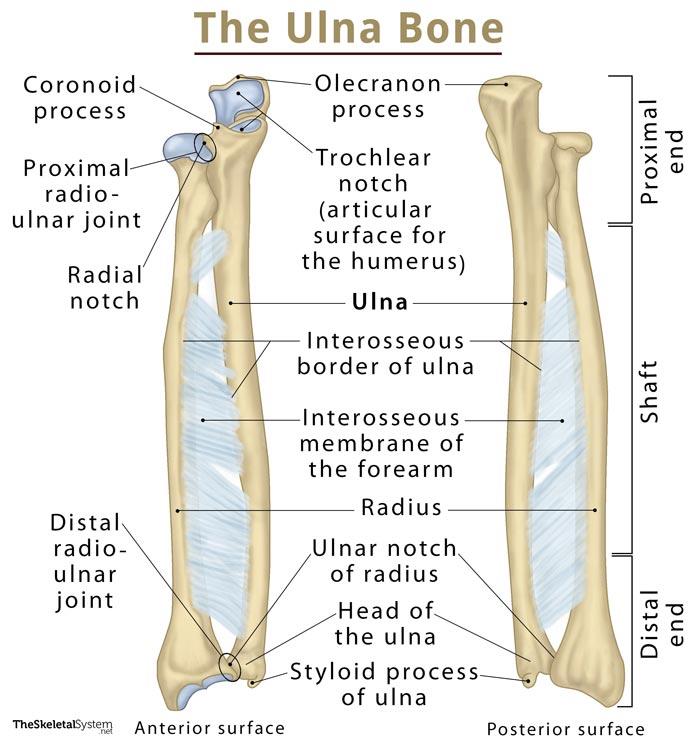
The long bone can be divided into three parts: the proximal or upper end, the shaft, and the distal or lower end [6].
The ulna articulates with only two bones, joining with the humerus and the proximal end of the radius on its proximal end, and the distal end of the radius on its distal end [2].
1. Proximal or Upper End
At the proximal end of ulna, there are four important bony landmarks, the olecranon process, coronoid process, trochlear notch, and the radial notch [7]. Another notable prominence is the tuberosity of ulna.
Elbow Joint: The head of the proximal ulna resembles a wrench, with a wide curved ‘c’ shaped surface formed by the trochlear or semilunar notch along with the olecranon process. The olecranon can be felt from outside as it forms the bony tip of our elbow [8]. The curved or crescent surface of the trochlear notch articulates with the trochlea of the humerus to form the hinge joint of the elbow. During extension of the arm, the olecranon inserts into the olecranon fossa, a deep recess or curve in the humerus, to keep the elbow from extending beyond its 180° range. The ulna is further secured in its place by the coronoid process (the distal end of the trochlear notch), protruding anteriorly to fit into the coronoid fossa, another smaller recess in the humerus, when the arm is flexed [3, 5].
Right below the coronoid process, the rough surface on the anterior side of the ulna meant for muscular attachments is known as the tuberosity of ulna [9].
Proximal Radio-Ulnar Joint: Lateral to the trochlear notch, at the end of the coronoid process, there is a small smooth surface called the radial notch that articulates with the proximal end of the radius to form the proximal radio-ulnar joint, so the radius can rotate around the ulna to maintain the flexibility of the elbow [5, 8].
| To identify the left/right ulna on a model, check which side the radial notch is located on while keeping the trochlear notch facing towards you. The radial notch is located on the same side as the hand, that is on the right side for the right hand, and on the left side for the left hand. |
2. Shaft
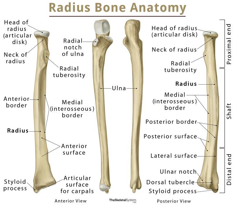
The shaft or body is the long middle part of the ulna bone. Moving down toward the distal side, the shaft tapers gradually [8] and has three prominent surfaces and three borders ― the anterior, posterior, interosseous borders and the anterior, posterior, medial surfaces [3].
The upper part of the shaft is somewhat pyramidal in shape, with a curve that makes it convex laterally and on the back [10]. There is a prominent ridge, known as the interosseous border of the ulna, running down the length of the lateral side of the shaft. This is where the interosseous membrane of the forearm, the thin fibrous sheet of tissue that holds the radius and ulna together, attaches to the ulna [5].
3. Distal or Lower End
Its distal end is much narrower compared to the proximal end, with two primary bony landmarks, the head of the ulna, and a styloid process [9].
The small rounded part on the distal ulnar surface is the head, and it articulates with the cup-shaped ulnar notch of the radius and the triangular fibrocartilage articular disc, a cartilage structure that keeps the ulna from forming any direct articulations with the carpal bones [7, 11]. The alignment of the ulnar head and the ulnar notch allows the distal end of the ulna work as a pivot so the radius can rotate around it in all directions [8].
The styloid process is a small bony protrusion extending from the posterior medial side of the head [7]. This is where the ulnar collateral ligament (UCL) of the wrist attaches [8].
Muscle and Ligament Attachments
The borders and surfaces of the ulnar shaft are the primary site for muscular attachments to this bone [3]:
Name of Muscle |
Insertion At Ulna |
| Triceps brachii | Olecranon process |
| Anconeus | Olecranon process |
| Brachialis | Olecranon process |
Name of Muscle |
Origin At Ulna |
| Pronator quadratus | Distal part of the anterior surface [9] |
| Supinator | Supinator crest of ulna (a prominent ridge running from the back of the radial notch to proximal lateral surface of the ulna) [13] |
| Flexor carpi ulnaris | Olecranon process and the posterior border of the ulnar shaft [12] |
| Flexor digitorum superficialis | Anterior and medial surfaces of the shaft |
| Flexor digitorum profundus | Anterior and medial surfaces of the shaft |
| Abductor pollicis longus | Posterior surface of the shaft |
| Extensor pollicis longus | Posterior surface of the shaft |
| Pronator teres | Coronoid process |
| Ponator quadratus | Distal part of the anterior surface |
| Extensor indicis | Distal part of the posterior surface of the shaft |
Blood Supply
Primary blood supply is provided by the ulnar artery, as well as its branch the common interosseous artery, which then further branches into the volar and posterior interosseous arteries, still supplying the ulna [6].
Nerve Distribution
The anterior interosseous nerve, branching from the median nerve, innervates the ulna on the volar side, while posteriorly it is innervated by the dorsal interosseous nerve, a branch of the radial nerve [6].
Primary Functions of the Ulna
- Forming the elbow joint with the humerus
- Shaping up and maintaining the structure of the forearm (along with the radius)
- Attaching with the vital muscles and ligaments of the lower arm and hand
- Working with the radius and carpal bones to move and rotate the wrist [2]
Proper movement of the ulna, along with all the attached muscles is instrumental for doing anything with our hand, from extending or flexing the arm, picking something up, throwing or holding something to eating, driving, and typing [8].
FAQ
Ulnar fractures are quite common, with the points where it joins with the radius and the fibrocartilage articular disc at the wrist being most frequently injured. Dislocations are also common, especially on the elbow side [14, 15]. Another condition associated with it is the Ulnar impaction syndrome (Ulnar abutment) where the ulna may be longer than the radius on the distal end, causing it to bump into the wrist bones, leading to pain [16].
The ulna is longer but much narrower than the radius [3]. Though early in life when a baby is only 4-5 months old, the ulna has a 50% larger diameter compared to the radius, it gradually reduces to become half of that of the latter as the person reaches adulthood [3].
The exact length of the ulna varies from one person to another, with research showing a possible correlation between the ulnar length and one’s height (the height of an individual may be estimated by calculating the length of his ulna bone) [4]. This is referred to as the ulna length-to-height formula.
References
- https://www.medicinenet.com/script/main/art.asp?articlekey=24396
- https://www.healthline.com/human-body-maps/ulna-bone
- https://www.kenhub.com/en/library/anatomy/the-radius-and-the-ulna
- https://www.ncbi.nlm.nih.gov/pubmed/28917221
- https://courses.lumenlearning.com/suny-ap1/chapter/bones-of-the-upper-limb/
- https://radiopaedia.org/articles/ulna
- https://www.earthslab.com/anatomy/ulna/
- http://www.innerbody.com/image_skelfov/skel21_new.html
- http://teachmeanatomy.info/upper-limb/bones/ulna/
- https://www.imaios.com/en/e-Anatomy/Anatomical-Parts/Shaft-of-ulna-Body-of-ulna
- http://teachmeanatomy.info/upper-limb/joints/wrist-joint/
- https://www.earthslab.com/anatomy/flexor-carpi-ulnaris/
- http://www.anatomyexpert.com/app/structure/1270/996/
- https://www.mayoclinic.org/diseases-conditions/dislocated-elbow/diagnosis-treatment/drc-20371692
- https://lifeinthefastlane.com/isolated-volar-distal-ulnar-dislocation/
- https://radiopaedia.org/articles/ulnar-impaction-syndrome