Scapula (Shoulder Blade)
What is Scapula
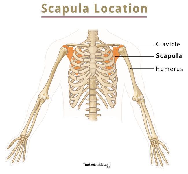
The scapula, alternatively known as the shoulder blade, is a thin, flat, roughly triangular-shaped bone placed on either side of the upper back. This bone, along with the clavicle and the manubrium of the sternum, composes the pectoral (shoulder) girdle, connecting the upper limb of the appendicular skeleton to the axial skeleton.
Where is the Scapula Located
The scapula is located on the upper back of the rib cage, extending from ribs 2-7. It lies between the upper arm bone, humerus, and collar bone or clavicle.
Scapula Facts
| Type | Flat bone |
|---|---|
| How many are there in the human body | 2 (1 on each side) |
| Articulates with | 1. Humerus at the glenohumeral or shoulder joint 2. Clavicle at the acromioclavicular joint |

How does the scapula move
The movement of this bone is coupled with the humerus, meaning whenever you move your arm, your scapula also moves. It can move in six different ways, towards (retract) and away (protract) from the vertebral column, up and down (elevate and depress), and also rotate upwards and downwards. There are 17 muscles attached to it that help to produce these movements.
Functions
The scapula helps in several daily movements and smooth motion of the upper arm, based on the movements mentioned above.
- It assists in both forward and backward movement of the pectoral girdle and chest muscles by moving closer and away from the vertebral column.
- During some motions, like shrugging the shoulders, the entire shoulder capsule moves up and down due to elevation and depression of this bone.
- It stabilizes the shoulder capsule during excessive arm motion by rotating upwards and downwards.
Development and Ossification
The scapula develops in the embryonic stage and ossifies from one primary and seven secondary centers. The primary ossification center appears near the glenoid cavity during the 8th week of fetal development. Out of seven, the first secondary center appears in the middle of the coracoid process during the first year and fuses by 15.
Another ossification center, called the subcoracoid center, develops in the root of the coracoid process around 10 and fuses by 16 to 18 years. Other centers, including one for the lower 2/3rds of the margin of the glenoid cavity, two for the acromion, one for the medial border, and one for the inferior angle, appear at puberty and fuse by the age of 25.
Anatomy – Parts of Scapula
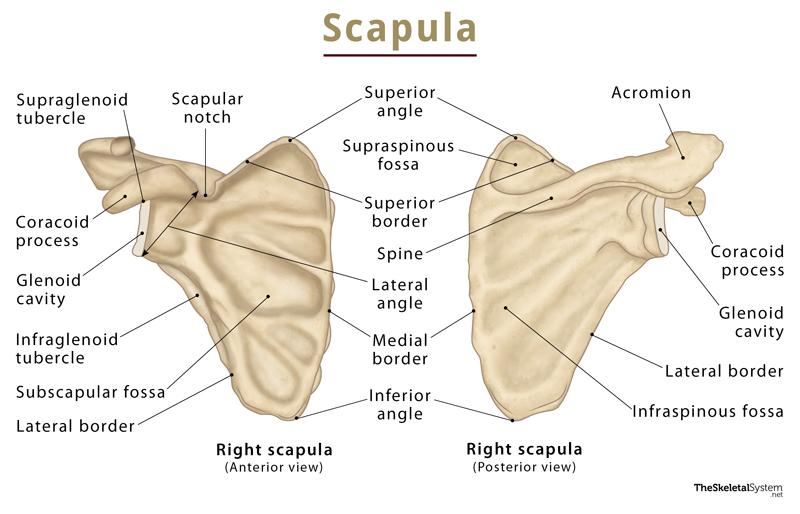
The body of the scapula consists of a triangular-shaped flat blade, with an apex pointed below. Since it is triangular, it bears three borders.
Borders and angles
- Superior border: It is the shortest and thinnest border.
- Medial border: It is a thin border running parallel to the vertebral column and is often referred to as the vertebral border.
- Lateral border: It is alternatively known as the axillary border, running towards the apex of the axilla. Out of the three borders, it is the thickest and strongest. It also bears the glenoid cavity, which articulates with the rounded head of the humerus, forming the shoulder joint or glenohumeral joint.
It also has three angles:
- Lateral angle: where the superior border converges with the lateral border.
- Superior angle: Where the superior border also meets with the medial border.
- Inferior angle: Where the medial and lateral borders meet.
Surfaces
1. Costal Surface
It is the anterior surface of the scapula facing the thoracic cage or ribcage.
It has a large concave depression over most of the surface, called the subscapular fossa, from where the rotator cuff muscle subscapularis originates.
This region is marked by longitudinal ridges, out of which a thick ridge joins the lateral border. This part of the bone acts as a lever for the action of the serratus anterior muscle, helping to move the arm away from the body.
A hook-like projection, called the coracoid process, originates from the superior border of the head of the scapula, projecting forward and curving laterally, lying underneath the clavicle.
2. Lateral Surface
This surface of the scapula faces the humerus.
Its important bony landmarks are:
Glenoid fossa – It is a shallow pyriform cavity located at the lateral angle of the scapula. It articulates with the rounded head of the humerus, forming the glenohumeral (shoulder) joint.
Supraglenoid tubercle – It is a small rough projection located immediately above the glenoid fossa near the base of the coracoid process.
Infraglenoid tubercle – It is a rough impression located on the lateral part of the scapula, immediately below the glenoid fossa.
3. Posterior Surface
This surface of the scapula faces outwards. Most of the rotator cuff muscles of the shoulder arise from here. Its important anatomical landmarks are:
Spine: It is a triangular plate of bone located on the posterior surface, running transversely across the scapula, dividing the dorsal surface of the scapula into supraspinous and infraspinous fossae. The two fossae remain connected by the spinoglenoid notch, situated lateral to the root of the spine, bridged by the spinoglenoid ligament. It has three borders and two surfaces. Its posterior border, the crest of the spine, bears upper and lower lips.
Supraspinous fossa: It is the area above the spine of the scapula. It is concave, smooth, and broader at its vertebral than at its humeral end. The supraspinatus muscle originates from the middle of this area. It is much smaller than the infraspinous fossa, bearing the spinoglenoid fossa on its side. The fossa houses the suprascapular canal, connecting the suprascapular notch and the spinoglenoid notch conveying the suprascapular nerve and vessels.
Infraspinous fossa: It is the area below the spine of the scapula. It is convex and much larger than the preceding one. At its upper part, towards the vertebral margin, it shows a shallow concavity. In the center, it is convex, while near the lateral border, it has a deep groove running from the upper toward the lower part.
Acromion: It is a large bony projection on the upper end of the scapula. It arches over the shoulder joint, articulating with the clavicle at the acromioclavicular (AC) joint.
Articulations
- Glenohumeral joint: This is a ball and socket joint formed between the glenoid fossa of the scapula and the rounded head of the humerus.
- Acromioclavicular joint: It is a gliding joint between the acromion of the scapula and the clavicle.
Muscles Attached to the Scapula
As the scapula has a large surface area, a large number of muscles get attached to it. The 17 muscles attached here fix the scapula to the thoracic wall and allow it to move. Four muscles, namely subscapularis, infraspinatus, teres minor, and supraspinatus, form the rotator cuff, covering the shoulder capsule.
These muscles are listed below and classified based on whether they originate from the scapula or insert into it.
Originating from the scapula
- Deltoid muscle: Originates from the lower border of the crest of the spine to the lateral border of the acromion. It helps to move the arm close and away from the body and also to rotate it at the shoulder joint.
- Supraspinatus muscle: Originates from supraspinous fossa. It sends the arm away from the body.
- Infraspinatus muscle: Originates from the infraspinous fossa and involves lateral rotation at the shoulder joint.
- Triceps brachii muscle (long head): Arises from the infraglenoid tubercle. It is responsible for elbow extension.
- Teres minor muscle: Originates from the lateral or axillary border of the posterior surface and performs lateral rotation at the shoulder joint.
- Teres major muscle: Arises from the posterior surface of the inferior angle and the lower part of the lateral border. It helps to bring the elbow towards the body and also to rotate at the shoulder joint.
- Latissimus dorsi muscle: Originating from the inferior angle, it performs several actions, such as protraction and retraction of the arm and medial rotation at the shoulder joint.
- Coracobrachialis muscle: Arises from the coracoid process. It is involved with retraction and depression at the shoulder joint.
- Biceps brachii muscle (long and short head): The long head originates from the supraglenoid tubercle, whereas the short head is from the coracoid process. It helps to bend the elbow.
- Subscapularis muscle: Originates from the subscapular fossa, performing depression and medial rotation at the shoulder joint.
- Omohyoid muscle: Arises from the superior border (adjacent to the suprascapular notch) and causes depression of the hyoid bone.
Inserting on the scapula
- Trapezius muscle: Gets inserted superiorly along the spine, acromion process, and clavicle. It helps to elevate and rotate the scapula during protraction of the humerus beyond 90 degrees.
- Levator scapulae muscle: Insert into the superior angle and medial border. They help in elevating the scapula.
- Rhomboid major muscle: Gets inserted in the medial border. It performs elevation and retraction of the scapula.
- Rhomboid minor muscle: Inserts above the scapular spine. Its actions include elevation and retraction of the scapula.
- Serratus anterior muscle: The insertion is along the medial border, from the superior to the inferior angle. It protracts, rotates, and stabilizes the scapula.
- Pectoralis minor muscle: Gets inserted into the coracoid process. It helps in the protraction and depression of the scapula.
Left and Right Scapula – How to Identify
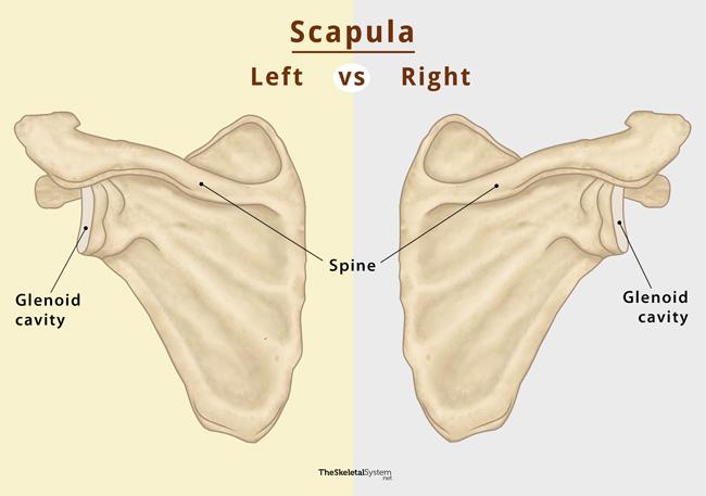
A quick way to identify whether the scapula comes from the right or left side of the body:
First, hold the bone at the inferior angle and orient it so that the convex posterior surface faces you. In this position, the glenoid cavity faces laterally outward, and the spine is clearly visible.
If the spine points at 2 o’clock, it is the right scapula. Alternatively, if it points at 10 o’clock, it is the left one.
Another way to identify the side is to observe which side the glenoid cavity is facing. While holding the bone in the position mentioned above, if the glenoid cavity faces right according to your body, that is the right scapula and vice versa.
FAQs
Ans. The muscles that stabilize the scapula are the serratus anterior, rhomboids, levator scapulae, and trapezius muscles.
Ans. No, the scapula is not a part of the axial skeleton.
Ans. Yes, the scapula is a part of the appendicular skeleton.
Ans. According to a research done on Europeans with Hispanic descent, it was found that female scapula is shorter than its male counterpart.
References
- The Scapula – Teachmeanatomy.info
- Scapula – Radiopaedia.org
- Scapula – Innerbody.com
- Scapula – Sciencedirect.com
- Scapula – Kenhub.com