Occipital Bone
Published on June 24th 2022 by staff
What is the Occipital Bone
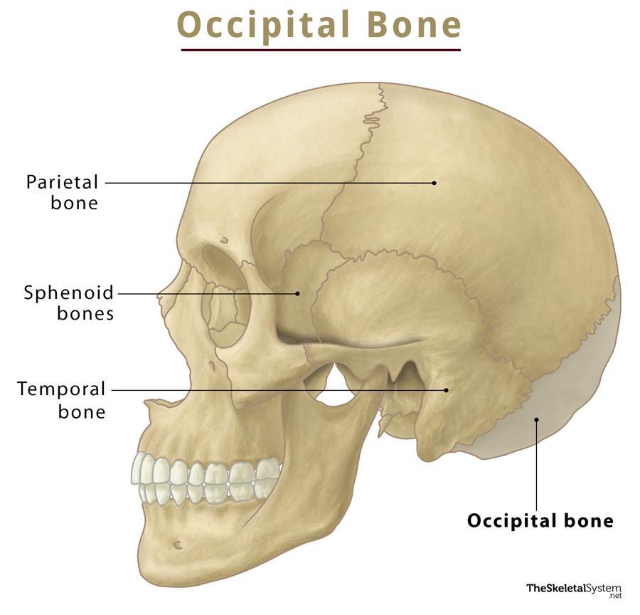
The occipital is an unpaired, trapezoidal cranial bone covering the back of the head. The curved bone resembles a shallow dish. It allows the spinal cord to pass from the brain into the spine.
Where is the Occipital Bone Located
It is the most posterior of all the cranial bones, forming the back of the head (occiput).
Quick Facts
| Type | Flat bone |
| How many are there in the human body | 1 |
| Articulates with | Total 6 bones: Parietal (paired), temporal (paired), Sphenoid, and atlas (C1) |
Function
It protects the occipital lobe and cerebellum of the brain, along with the associated nerves and blood vessels.
Occipital Bone Anatomy
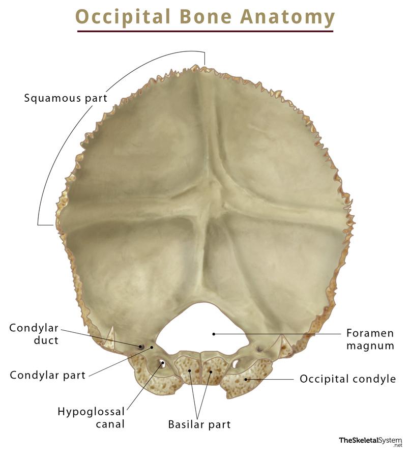
This trapezoidal-shaped bone is externally convex and internally concave. It can be divided into the basilar, two condylar/lateral, and the squamous parts. Each part has two surfaces: upper or external and lower or internal.
Surfaces and Landmarks
Basilar Part
This quadrilateral part of the bone is adjacent to the petrous part of the temporal bone and anterior to the foramen magnum. It has an upper surface and a lower surface.
During adolescence, the upper surface of the basilar part articulates with the sphenoid bone to form the clivus. It has a broad, shallow groove that supports the medulla oblongata. The lower surface features the pharyngeal tubercle, where the superior pharyngeal constrictor muscle and fibrous pharyngeal raphe insert. The other muscles attached to the lower surface are rectus capitis anterior and longus capitis.
Condylar Parts
The condylar parts are commonly known as the lateral parts of the occipital bone, as they are found lateral to the foramen magnum. Each of the two condylar parts contains an upper and lower surface. It features two kidney-shaped prominences called occipital condyles that form articulation with the first cervical vertebra (C1), thus giving rise to the atlanto-occipital joint.
The condylar canals are located just behind the condyles, through which the condylar emissary veins pass. The canals also connect the external vertebral venous plexuses with the sigmoid sinuses. The hypoglossal canal lies on the inferior surface of the condylar part through which the hypoglossal nerve leaves the cranium.
Squamous Part
It is the largest of all four parts and contains internal and external surfaces. This part features the external occipital protuberance, a bony prominence in the middle of the outer surface.
The trapezius muscle attaches here. The external surface also has three curved lines, called the nuchal lines. They are:
- Supreme nuchal line: Laterally extends from the external occipital protuberance. The epicranius muscle and epicranial aponeurosis originate from here.
- Superior nuchal line: Running inferior to the squamous part, it serves as a site of origin for the trapezius, splenius capitis, and sternocleidomastoid muscles.
- Inferior nuchal line: Found further inferior to the superior nuchal line, this is where the semispinalis capitis muscle gets inserted.
Several grooves mark the internal surface of this part by dural venous cranial sinuses, such as the superior sagittal sinus, the transverse sinuses, and the sigmoid sinus. The groove above the transverse sinus houses the occipital lobes and cerebellum of the brain.
Foramen Magnum
All the above four parts are arranged around a large opening at the back of the bone, called the foramen magnum. It allows passage to the spinal cord. Specific structures that pass through the foramen magnum are the brain stem (medulla oblongata), a spinal branch of the accessory nerve, anterior and posterior spinal arteries, the vertebral arteries, the tectorial membrane, and alar ligaments.
Borders and Articulations
The bone articulates with total 6 bones. Among them, 2 are paired (parietal and temporal), while the other 2 are unpaired (sphenoid and C1/first cervical vertebrae). The occipital is the only cranial bone to form an articulation with a spine bone.
- Lambdoid Suture: Between the occipital and parietal bones.
- Occipitomastoid Suture: Between the occipital and the mastoid part of the temporal bone.
- Petro-Occipital Suture: Between the occipital bone and the petrous part of the temporal bone.
- Spheno-Occipital Suture: Btween the occipital and sphenoid bones. It gradually disappears as the two bones fuse during adolescence.
- Atlanto-Occipital Joint: Joint formed by the occipital bone with the first cervical vertebrae (C1). It is the only joint between a cranial bone and a vertebra.
Development and Ossification
The ossification of this bone starts around the 9th week of fetal life. The squamous part of the bone undergoes membranous ossification, whereas its other parts have cartilaginous ossification.
The four parts remain separate at birth but fuse as a child grows up, with the squamous and condylar parts fusing at around 2 years, while the condylar and basilar parts fuse by 6 years.
References
- Occipital bone — Kenhub.com
- Anatomy, Head and Neck, Occipital Bone, Artery, Vein, and Nerve — Ncbi.nlm.nih.gov
- Occipital bone — Radiopaedia.org
- Occipital Bone Anatomy — Getbodysmart.com
- Occipital Bone — Sciencedirect.com