Parietal Bone
Published on November 14th 2022 by staff
What is the Parietal Bone
The parietal is a large, flat, paired cranial bone that forms a considerable portion of the skull.
Where is the Parietal Bone Located
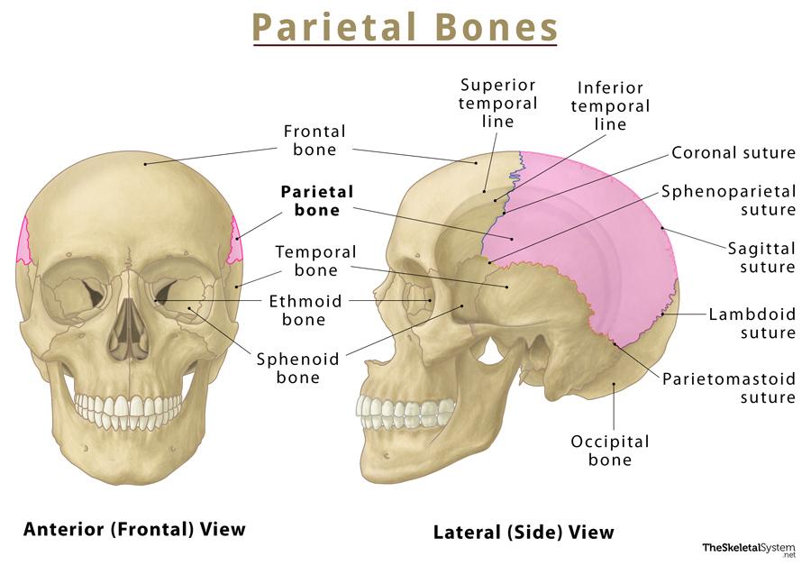
The parietal bones are located on either side of the skull, making up most of the top and sides of the head. They are part of the neurocranium, along with frontal, occipital, sphenoid, temporal, and ethmoid bones, sharing borders with the first 4 of these 5 bones. The name ‘parietal’ derives from their location over the brain’s parietal lobes.
Quick Facts
| Type | Flat bone |
| How many are there in the human body | 2 |
| Articulates with | Frontal (unpaired), occipital (unpaired), sphenoid (unpaired), and temporal (paired) |
Functions
Its primary role as a skull bone is structural. The paired bones have the following functions:
- Forms the sides and roof of the skull towards the back, hence shaping the head.
- The left and right parietal bones protect the brain’s left and right parietal lobes, respectively.
- Limits any swelling in case of a brain condition involving an infection, bleeding, or an increase in cerebrospinal fluid levels.
Parietal Bone Anatomy and Landmarks
The curved bone is somewhat quadrilateral, with 2 rather broad surfaces and 4 borders.
Borders and Sutures
The borders articulate with the other bones around the parietal, forming 5 sutures. Sutures are immovable joints in the skull.
- Sagittal Border: It is the upper medial border, where the thickest and longest suture forms. This is where the left and right parietal bones join with each other, forming the sagittal suture.
- Squamosal Border: The lower border, where the parietal and the greater wing of the sphenoid bones join, giving rise to the sphenoparietal suture. It also joins with the temporal bone at the parietomastoid suture. The border is thin at its starting point, arching slightly along the middle, then thickening towards the back.
- Frontal Border: The most serrated of all the parietal bone’s margins, this is where the bone articulates with the posterior part of the frontal bone. The associated suture is called the coronal suture.
- Occipital Border: This is where the bone articulates with the occipital bone at the back. Also highly serrated, it forms the lambdoid suture.
Angles
Since there are 4 borders, there are 4 angles where the borders meet:
- Frontal Angle: Where the sagittal border meets the frontal border.
- Sphenoidal Angle: The thin, pointed angle where the frontal border intersects the squamosal.
- Occipital Angle: The angle at the upper back side of the bone; it is formed where the sagittal and occipital borders meet. It is the most rounded of all the angles in the parietal bone.
- Mastoid Angle: The angle at the lower back side, it forms where the occipital border intersects the squamosal border.
Surfaces
The External Surface
As the name suggests, this is the convex outer surface of the bone that protects the brain. This smooth curved surface also shapes the back of the head. It has several important landmarks for muscular attachments:
- The superior temporal line is the arch between the occipital and frontal borders; it is where the superficial temporal fascia attaches to the parietal.
- The inferior temporal line is where the temporalis muscle originates. This line forms a similar arch to the superior temporal line but is located lower in the skull.
- Parietal foramina are present at the upper back side of each of the parietal bones, near the sagittal border. These foramina drain into the superior sagittal sinus and allow passage to the occipital artery’s branches.
- The parietal eminence is the central part of the parietal bones, where the bones start to ossify.
The Internal Surface
It is the concave inner surface of the bone on the side of the brain. The internal surface is highly irregular to accommodate different blood vessels. The most important of them include the following:
- The arterial grooves, or grooves for the middle meningeal artery
- The grooves for superior sagittal sinus
- The grooves for sigmoid sinus
The granular foveolae surrounding the groove for superior sagittal sinus and containing arachnoid granulations
References
- Parietal Bone: KenHub.com
- Parietal Bone: RadioPaedia.org
- Parietal Bone Anatomy & Function: Study.com
- Parietal Bone: Anatomy.app
- Parietal Bone Anatomy: GetBodySmart.com