Palatine Bone
Published on June 24th 2022 by staff
What is the Palatine Bone
Palatine is one of the eight facial bones that form the facial skeleton or viscerocranium, along with others. This paired, irregular, L-shaped bone forms a portion of the hard palate, besides the nasal cavity and orbits.
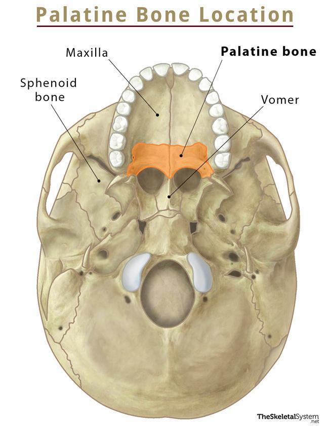
Where is the Palatine Bone Located
The bone is located between the maxillae and sphenoid bone, specifically between the palatine process of the maxillae and the pterygoid process of the sphenoid bone.
Quick Facts
| Type | Irregular bone |
| How many are there in the human body | 2 |
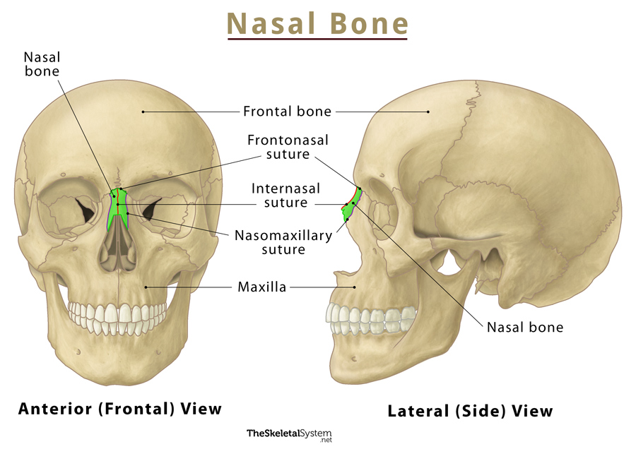
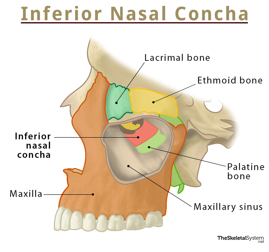
| Articulates with | 5 bones: maxilla, vomer, inferior nasal concha, ethmoid, and sphenoid bones. |
Functions
- It forms a part of the hard palate by articulating with the palatine process of the maxilla. This palate acts as the roof of the oral cavity and the floor of the nasal cavity.
- The palatine also forms the floor of the orbital cavity along with the 6 other orbit bones.
- The bone, along with the palatine process of the maxilla, gives rise to the lateral walls of the nasal cavity.
Anatomy
This bone is composed of two plates, horizontal and perpendicular, that connect at nearly right angles, giving the bone its characteristic L-shape. It also contains three processes: pyramidal, orbital and sphenoidal.
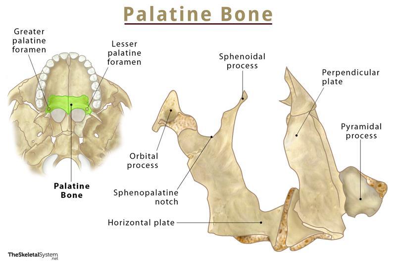
Horizontal Plate
As the name implies, this quadrangular plate is the horizontal part of the bone. Thus it forms the baseline of the L-shaped bone. It forms a part of the hard palate and nasal cavity. The horizontal plate features four borders (medial, lateral, anterior, and posterior) and two surfaces (nasal and palatine).
Borders
- Medial Border: The left and right palatine bones join at their respective medial border, forming a median palatine suture.
- Lateral Border: This border is a continuation of the perpendicular plate, having the greater palatine foramen.
- Anterior Border: The horizontal plate’s anterior border articulates with the maxilla’s palatine process, jointly forming the hard palate.
- Posterior Border: It faces the back wall of the pharynx.
Surfaces
- Nasal surface: As the name suggests, it partially makes up the posterior part of the nasal cavity’s floor and inferior nasal meatus.
- Palatine surface: As the name implies, it forms a part of the hard palate. The surface contains two important foramina:
- Greater palatine foramen: It is a small opening that allows the passage of greater and lesser palatine nerves running from the greater palatine canal and reaching the oral cavity. Along with these nerves, the descending palatine vessels run through this opening. The canal passes through the palatine and sphenoid bones, connecting the pterygopalatine fossa to the oral cavity. At the terminal, the canal gives rise to the lesser palatine canal.
- Lesser palatine foramina: This foramen leads to the lesser palatine canal, carrying the lesser palatine vessels and nerves.
Perpendicular Plate
The perpendicular plate rises at a nearly 90° angle with the lateral border of the horizontal plate, giving the bone its characteristic L-shape. It is composed of several borders and surfaces.
Borders
Its 4 borders articulate with the adjacent bones and give rise to some essential bony landmarks.
- Anterior border: It has a laminar projection to articulate with the inferior nasal concha, partially forming the maxillary sinus’ medial wall.
- Posterior border: This serrated border articulates with the sphenoid bone’s medial pterygoid plate.
- Superior border: It articulates with the body of the sphenoid bone. The border features a deep notch, the sphenopalatine notch, between its orbital and sphenoidal processes. It remains enclosed by the sphenoid body, leading to the formation of the sphenopalatine foramen. This foramen connects the pterygopalatine fossa with the nasal cavity.
- Inferior border: This border is a continuation of the lateral border of the horizontal plate.
Surfaces
It features two surfaces:
- Nasal surface: This partially forms the lateral wall of the nasal cavity, particularly the superoposterior part of the nasal septum. On its superior side, it articulates with the cribriform plate of the ethmoid bone.
- Maxillary surface: This mostly rough and irregular surface articulates with the nasal surface of the maxilla.
Processes
The bone features three main processes: pyramidal, orbital, and sphenoidal.
1. Pyramidal process: It emerges from the junction of horizontal and perpendicular plates, running between the sphenoid bone’s medial and lateral pterygoid plates. Its posterior surface joins with the pterygoid plates of the sphenoid bone to form a part of the pterygoid fossa.
2. Orbital process: This process originates anteriorly from the perpendicular plate. It features three articular, two non-articular surfaces, and a narrow neck.
Articular surfaces
- Anterior or maxillary surface: It articulates with the maxilla.
- Posterior or sphenoidal surface: It articulates with the sphenoid bone.
- Medial or ethmoidal surface: It articulates with the ethmoid bone.
Non-articular surfaces
- Superior or orbital surface: It partially forms the floor of the eye cavity.
- Lateral surface: It also partially forms the pterygopalatine fossa.
3. Sphenoidal process: It continues from the upper part of the posterior border, forming a part of the pterygopalatine fossa. The medial border of this process forms an articulation with the ala of the vomer.
Muscle Attachments
The four muscles that attach to this bone are:
- Medial pterygoid muscle: It attaches to the pyramidal process
- Superior pharyngeal constrictor muscle: This muscle gets attached to the horizontal plate
- Tensor palate: This one attaches to the horizontal plate as well
- Uvula muscle: Another muscle that gets attached to the horizontal plate
Ossification
The bone undergoes intramembranous ossification from a single center, which appears around the 6th–8th week of fetal life. The center first emerges at the junctional point of horizontal and perpendicular plates.
References
- Palatine bone – Kenhub.com
- Palatine bone – Radiopaedia.org
- Palatine Bone – Sciencedirect.com
- Palatine Bone Anatomy – Getbodysmart.com