Vomer
Table of Contents:
Published on June 24th 2022 by staff
What is the Vomer
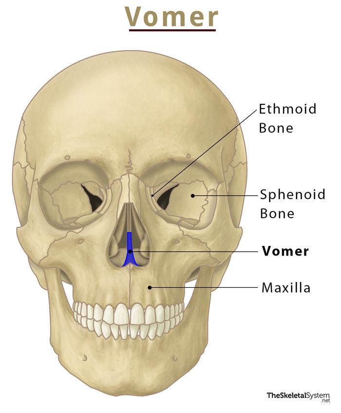
The vomer is one of the fourteen facial bones that form the facial skeleton or viscerocranium. This thin, flat, unpaired bone sits in the center of the nasal cavity, constructing the nasal septum. The bone is named so due to its shape; as in Latin, the word vomer refers to ‘plowshare’.
Where is the Vomer Bone Located
When looking at a skull from the front, this bone can be seen located vertically along the nasal cavity.
Quick Facts
| Type | Irregular bone |
| How many are there in the human body | 2 |
| Articulates with | 5 bones: maxilla, vomer, inferior nasal concha, ethmoid, and sphenoid bones. |
Functions
- Plays an important role in dividing the nasal cavity as it forms the lower back part of the nasal septum along with the septal cartilage and the ethmoid bone.
- Also features multiple grooves through which several nerves and blood vessels of the nasal cavity pass.
Anatomy
Surfaces
As the vomer is a flat bone, it has two surfaces. It bears an oblique groove called the nasopalatine or vomerine groove on each surface. The nasopalatine nerves and vessels pass through this groove.
Its vertical perpendicular plate is the most important structure, forming the majority of the bone. The vomer projects upward horizontally to form the wings or ala of the vomer.
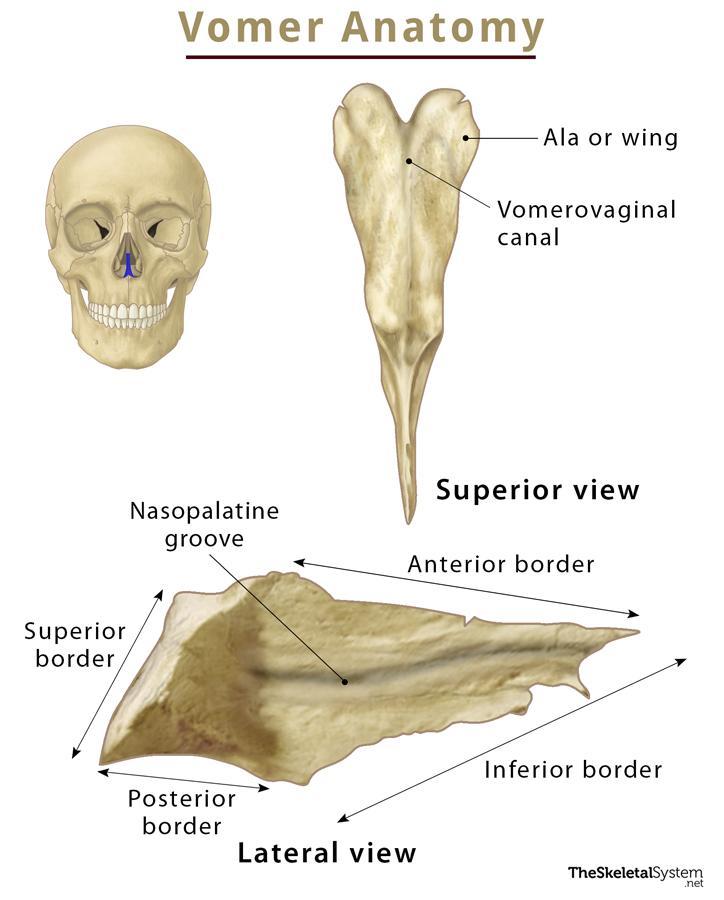
Borders
The four borders of this trapezoid-shaped bone are:
- Superior border: It is the thickest border, featuring two lateral wing-like projections, the alae of the vomer. In between the two alae, a deep furrow is present where the rostrum of the sphenoid articulates. The margins of alae articulate with the vaginal process of the sphenoid and the sphenoidal process of the palatine bone. The vomerovaginal canal runs between these alae and the vaginal process of the sphenoid bone.
- Inferior border: It articulates with the median nasal crest, jointly formed by the maxillae and palatine bones.
- Anterior border: It is the longest border of the vomer, with its upper half articulating with the ethmoid bone’s perpendicular plate. On the other hand, its grooved lower half joins with the inferior margin of nasal septal cartilage.
- Posterior border: This concave border does not articulate with any other neighboring bones. It is thick and bifid on the upper side, becoming thinner as we move down. Its only job is to separate the posterior nasal apertures or conchae (internal nostrils).
Ossification
Vomer undergoes intramembranous ossification from two centers during the 9th week of the fetal stage. The centers develop in the mucoperichondrium at the lower border of the nasal septum.
References
- Vomer — Kenhub.com
- Vomer — Sciencedirect.com
- Vomer — Radiopaedia.org
- Vomer — Anatomystandard.com