Cuneiform Bones
What are the Cuneiform Bones
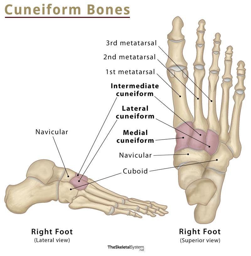
Cuneiforms are a set of three bones present in the midfoot. In Latin, ‘cuneus‘ means ‘wedge’, and ‘form’ refers to the shape. For this reason, these tarsal bones are named so due to their wedge shape.
From medial to lateral, these bones are named as:
- Medial Cuneiform
- Intermediate Cuneiform
- Lateral Cuneiform
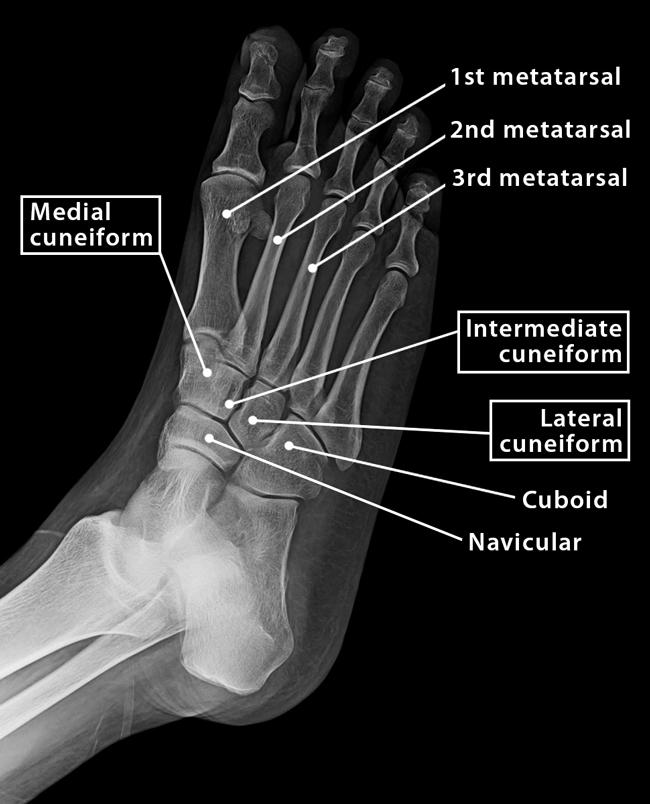
Where are the Three Cuneiform Bones Located in the Foot
The cuneiforms are located medially on the midfoot. They are positioned medial to the cuboid and between the navicular and the first three metatarsal bones.
Cuneiform Bone Facts
| Type | Short bone |
| Number in the human body | 6 (3 cuneiforms in each foot) |
Functions
The cuneiform bones help to form the transverse arch of the foot, which aids in maintaining body balance while standing or walking.
Structure and Anatomy of the Cuneiform Bones
1. Medial Cuneiform
Also known as the first cuneiform, it is the largest of the three cuneiforms. It is located at the medial side of the foot, anterior to the navicular bone, posterior to the first metatarsal, and lateral to the intermediate cuneiform. It exhibits multiple facets separated by bony ridges, articulating with the adjacent bones.
Bony Landmarks
The medial surface is rough, large, square, and subcutaneous. So, it is easily palpable.
The distal surface features a large kidney-shaped facet for the attachment of the first metatarsal bone.
The proximal surface bears a pyriform facet for the navicular.
The lateral surface remains marked by an inverted L-shaped facet along the posterior and superior margins for the intermediate cuneiform bone. A vertical ridge separates the anterosuperior part of the facet. The second metatarsal bone gets attached here.
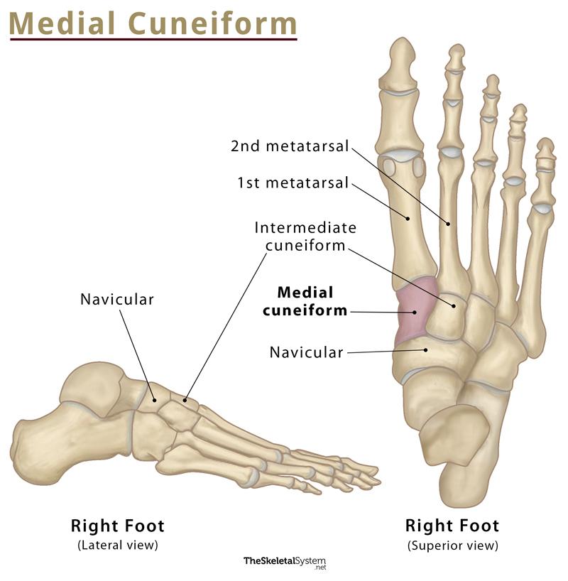
Articulations
The medial cuneiform bone articulates with four bones:
- Navicular
- Intermediate cuneiform
- First metatarsal
- Second metatarsal
Muscle Attachments
Two muscles, tibialis anterior and peroneus longus get inserted at the medial cuneiform bone.
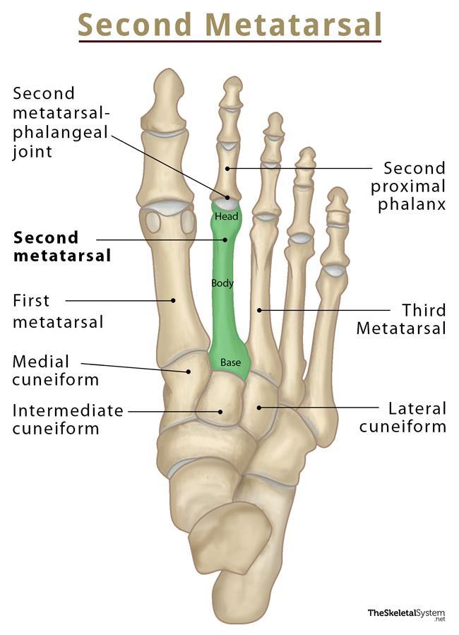
2. Intermediate Cuneiform
It is also known as second cuneiform or middle cuneiform, as it situated between the other two. This is the smallest of the three.
Bony Landmarks
The proximal and distal surfaces bear triangular articular facets for the second metatarsal bone and navicular, respectively.
Its medial surface partly articulates with the medial cuneiform.
The lateral surface bears a vertical facet along its posterior margin that articulates with the lateral cuneiform.
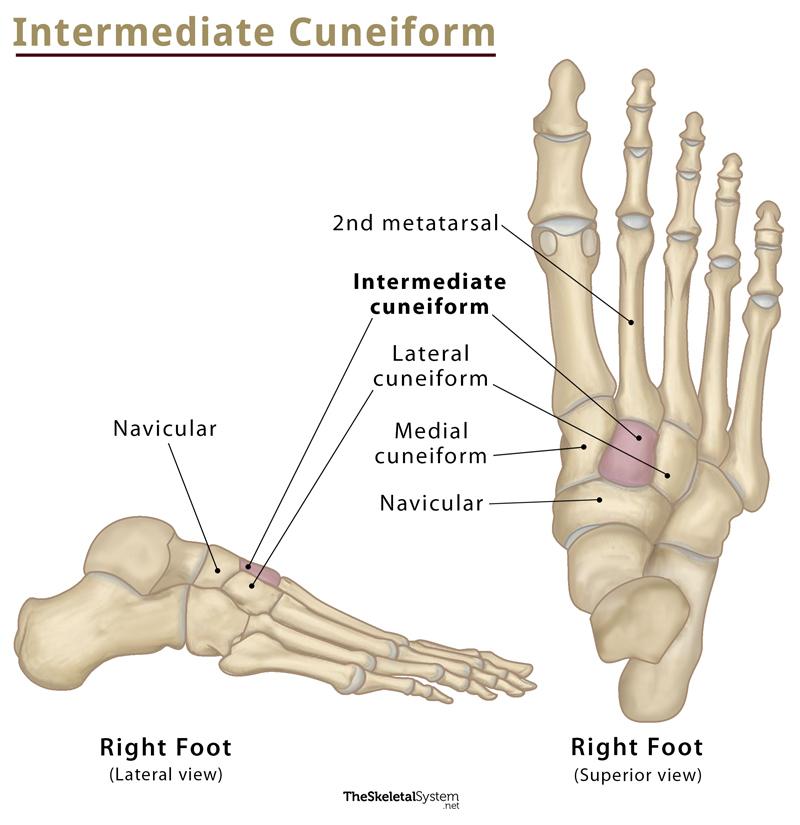
Articulations
It articulates with four bones:
- Navicular
- Medial cuneiform
- Lateral cuneiform
- Second metatarsal
Muscle Attachments
Its plantar surface receives the tibialis posterior tendon.
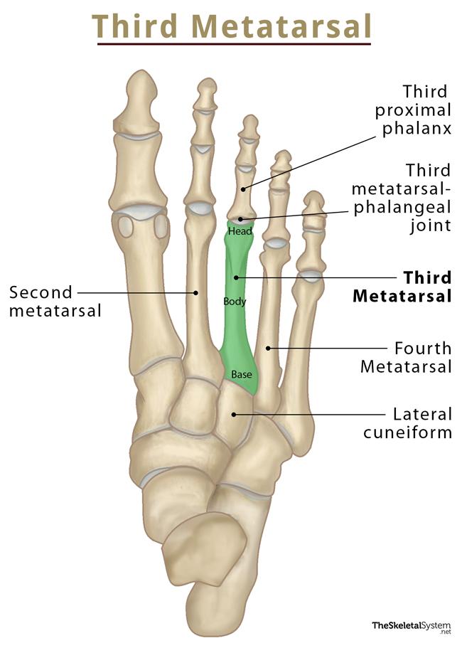
3. Lateral Cuneiform
The lateral cuneiform bone, also known as the third or external cuneiform, is medium-sized compared to the other two.
Bony Landmarks
The proximal surface is rough in its lower one-third and has a triangular facet in its upper two-thirds for the navicular bone.
A triangular or oval facet marks the lateral surface for the cuboid. It also features an articular surface on the medial side for the attachment of intermediate cuneiform.

Articulations
The lateral cuneiform bone articulates with four bones:
- Intermediate cuneiform
- Cuboid
- Navicular
- Third metatarsal
Muscle Attachments
Two muscles get attached to the lateral cuneiform bone. The tibialis posterior inserts into the bone, while the flexor hallucis brevis originates from it.
References
- Cuneiform bones – Kenhub.com
- Cuneiform bones – Radiopaedia.org
- Cuneiform bones – Anatomy.net
- Cuneiform bones – Earthslab.com