Hyoid Bone
Published on April 23rd 2022 by staff
What is the Hyoid Bone
The hyoid, also known as the lingual or tongue bone, is a small horseshoe-shaped bone in the neck. The name of this bone has been derived from the Greek word ‘hyoeides’, meaning ‘upsilon-shaped’ or ‘U-shaped’.
It is one of the most unique bones in the human body as it does not directly articulate with any other bone, having only muscular, ligamentous, and cartilaginous attachments. Due to this, it is referred to as a free-floating or sesamoid bone.
Where is the Hyoid Bone Located
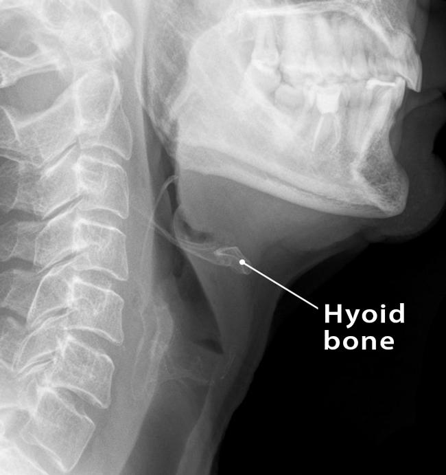
The bone is located in the mid-neck region, between the mandible and the thyroid cartilage, anterior to the pharynx and epiglottis at the level of the third cervical vertebra (C3). It remains suspended from the tips of the styloid processes of the temporal bones by the stylohyoid ligaments.
The hyoid can be easily palpated upon extension of the neck.
Quick Facts
| Type | Sesamoid bone |
| How many are there in the human body | 1 |
| Articulates with | None |
Functions
- Serves as the attachment point for the tongue and other muscles in the oral cavity floor.
- Aids in a wide range of muscle activities required for speaking and swallowing.
- Protects the esophagus.
Hyoid Bone Anatomy and Landmarks
This horseshoe-shaped bone comprises a body, two greater horns (cornua), and two lesser horns (cornua). The body of the hyoid is positioned centrally, while the horns or cornua are on both sides.
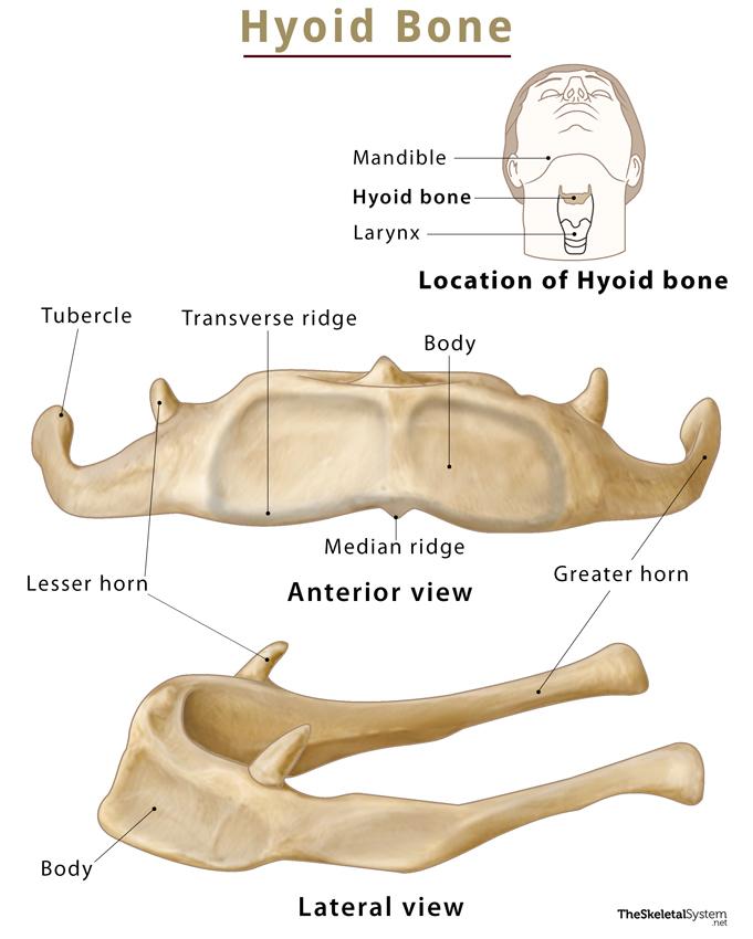
Body
This part of the bone stretches laterally, forming the central, irregular, quadrilateral-shaped, broad segment. It forms the convexity of the distinctive U-shape of the bone with its convex anterior and concave posterior surfaces. The body features a vertical median ridge in the middle, projecting inferiorly. Apart from this, the upper half of the body is also marked by transverse ridges with a slight downward convexity. On both extremities, it gets fused with the hyoid horns or cornua.
Greater Horns (Cornua)
These long, thin structures project from each end of the body, extending posterolaterally. The horns are wide in front and become narrow as they extend backward. The tip of each horn slightly expands to form a tubercle, where the lateral thyrohyoid ligament attaches.
Lesser Horns (Cornua)
The lesser horns are two small, conical bony protrusions, situated in the line of the transverse ridge on the body, and are a continuation of the same. It arises from the superior aspect of the hyoid bone, at the junction of the body and greater horn, projecting superoposteriorly towards the styloid process of the temporal bone, where the stylohyoid ligament attaches.
Articulations
The hyoid bone does not articulate with any other bone directly. Instead, it is connected to adjacent bones by muscular and ligamentous attachments.
Attachments
Here is a list of the muscles and ligaments that the hyoid bone attaches to.
Muscles
Several muscles originate from and insert into the hyoid bone. They are as follows:
I. Muscles originating from the hyoid bone
1. Hyoglossus Muscle: It originates from the anterior part of the body and helps to depress the tongue.
2. Middle Pharyngeal Constrictor: It originates from the bone and is responsible for constricting the middle part of the pharynx during swallowing.
II. Muscles inserting into the hyoid bone
Muscles that insert on the upper surface of the bone are known as suprahyoid muscles, while those attached to the lower surface are called infrahyoid muscles.
A. Suprahyoid Muscles
All these muscles help in elevating the hyoid.
1. Digastric Muscle: This muscle travels anteroinferiorly to get inserted at the junction of the greater cornu and body. It helps to depress and retract the chin and facilitate the opening of the mouth.
2. Mylohyoid Muscle: It gets inserted into the anterior part of the body and raises the hyoid bone during swallowing.
3. Geniohyoid Muscle: This muscle gets inserted on the anterior surface of the body. It acts by protracting and elevating the hyoid during swallowing.
4. Stylohyoid Muscle: It gets inserted at the junction of the greater cornu and body. This muscle retracts and elevates the bone during swallowing.
B. Infrahyoid Muscles
All of these muscles depress the bone.
1. Sternohyoid Muscle: The muscle gets inserted in the inferior surface of the hyoid bone.
2. Omohyoid Muscle: It inserts on the inferolateral surface of the hyoid.
3. Sternothyroid muscle: This muscle does not directly attach to the hyoid bone. It inserts on the oblique line of the thyroid cartilage.
4. Thyrohyoid muscle: It is the continuation of the sternothyroid, inserting into the body of the hyoid and greater cornu.
5. Genioglossus Muscle: It gets inserted into the body of the hyoid bone.
Ligaments
1. Stylohyoid Ligament: The ligament extends from the styloid process of the temporal bone to the lesser horn of the hyoid bone.
2. Hyoepiglottic Ligament: It attaches to the body and greater cornu of the hyoid bone, connecting the bone to the anterior aspect of the epiglottis.
Membranes
Thyrohyoid Membrane: It is an extrinsic membrane that originates from the superior border of the thyroid cartilage and attaches to the posterior surface of the body and the greater horns. This membrane anchors the laryngeal skeleton to the hyoid bone.
Development and Ossification
It is ossified from six centers: two for the body and one for each cornu.
FAQs
Ans. The hyoid bone is a part of the axial skeleton.
Ans. The hyoid bone fracture is very rare, almost 0.002% of all fractures in humans. The most common cause of hyoid bone fracture is neck strangulation, which majorly happens during homicides. So, a person generally does not die due to the fracture but rather the choking that leads to oxygen deprivation.
References
- The Hyoid Bone — Teachmeanatomy.info
- Hyoid bone — Kenhub.com
- Anatomy, Head and Neck, Hyoid Bone — Ncbi.nlm.nih.gov
- Hyoid bone — Radiopaedia.org
- The Hyoid Bone — Courses.lumenlearning.com