Appendicular Skeleton
What is the Appendicular Skeleton
The appendicular skeleton is one of the two major groups of bones in the human skeleton. It consists of the bones of the limbs (or appendages), and the bones that attach the limbs to the rest of the body.
It includes a total of 126 bones, including those in the arms, legs, and shoulder and pelvic girdle bones.
Basic Function of the Bones in the Appendicular Skeleton
The primary function of the appendicular skeleton is to provide shape and structure to the limbs, allowing them to work properly.
Parts of the Appendicular Skeleton With Names of Bones
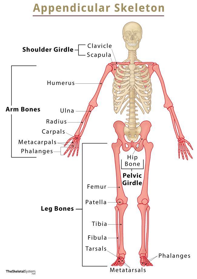
Shoulder (Pectoral) Girdle
- Clavicle (2): The thin bones positioned horizontally in the shoulder region articulate with the scapula on one side and the sternum (axial skeleton) on the other.
- Scapula (2): The flat, triangular bones that can easily be felt on both sides of our back. The wide bones support several muscles in the shoulder joint. The scapula articulates with the clavicle and the humerus — the upper arm bone.
Arm Bones (Upper Limb Bones)
There are 30 bones on each arm.
- Humerus (2): The longest and only bone in the upper arm, between the shoulder and elbow. The humerus articulates with the scapula on its proximal end, and the lower arm bones, radius, and ulna, on its distal end, connecting the shoulder to the elbow.
- Radius (2): Relatively thicker of the two lower arm bones, the radius is the one located on the thumb’s side, between the elbow and wrist.
- Ulna (2): Longer than the radius, it is located on the side of the little finger. The radius and ulna connect the elbow to the wrist, allowing us to rotate the forearm and wrist.
- Carpals (16): The 8 irregular bones lying on the wrist in each hand. These bones articulate with each other, as well as to the radius, ulna, and metacarpals.
- Metacarpals (10): The 5 long bones in each hand forming the distal part of the palm. These articulate with the lowermost (proximal) row of phalanges.
- Phalanges (28): The 14 bones that form the fingers; each finger has 3 phalanges, except the thumb, which has 2.
Pelvic Girdle
- Hip Bone: It is one large bone formed of three irregular bones that fuse during puberty and early adulthood.
The pelvic girdle is responsible for bearing the body’s weight, allowing us to stand/sit straight and walk. So, it has some strong ligaments that attach it to the axial skeleton. The hip bone articulates with the femur, as well as the sacrum (through ilium), which is an axial skeleton bone. So, it connects the legs to the axial skeleton.
Leg Bones (Lower Limb Bones)
- Femur (2): The heaviest, longest, and strongest bone in the human body, the femur is the only bone in the upper leg or thigh region. On its proximal end, it articulates with the hip bone, while its distal end articulates with the tibia and patella to form the knee joint.
- Tibia (2): The bigger, and only weight-bearing bone in the lower leg, it articulates with the femur on one end and the tarsal bones on the other, thus connecting the foot to the upper leg.
- Fibula (2): The smaller of the two lower leg bones, it is an attachment point for multiple leg muscles. The fibula articulates with the tibia at both its proximal and distal ends.
- Patella (2): Also known as the kneecap, it is the biggest sesamoid bone in the body, forming the knee. There is no corresponding bone for the patella in the arm. It stays embedded in the quadriceps femoris muscle’s tendons.
- Tarsals (14): Similar to the carpals, the tarsals are the small irregular bones forming the heels and part of the arch. Unlike carpals, there are only 7 tarsal bones in each foot.
- Metatarsals (10): These are the bones corresponding to the metacarpals in hand, and like them, these articulate with the toe bones, forming the distal part of the foot’s arch.
- Phalanges (28): Often referred to as the phalanges of the foot, these are the bones forming the toes. Each toe has 3 phalanges, except the big toe, which has 2.
An easy way to memorize the appendicular skeleton bones is that these are located in body regions we have in pairs of left and right — the shoulder, arms, pelvic girdle, and legs. These are the bones of the appendages. Whereas the axial skeleton includes body regions like the skull and spine, which are located centrally.
References
- Human Appendicular Skeleton – Lumenlearning.com
- Appendicular Skeleton (126 bones) – Training.seer.cancer.gov
- Types of Skeletal Systems – Human Appendicular Skeleton – Bio.libretexts.org
- Lesson Explainer: The Appendicular Skeleton – Nagwa.com
- Anatomy, Appendicular Skeleton – Ncbi.nlm.nih.gov/books.
- Appendicular Skeleton: Function & Anatomy – Study.com