Temporal Bones
Published on March 31st 2022 by staff
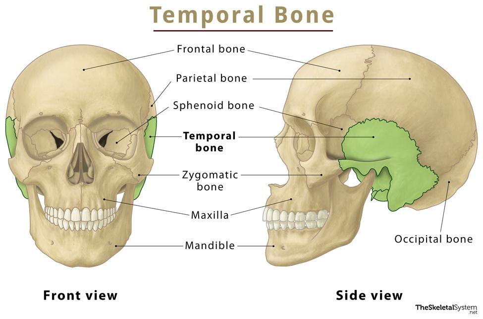
What is the Temporal Bone
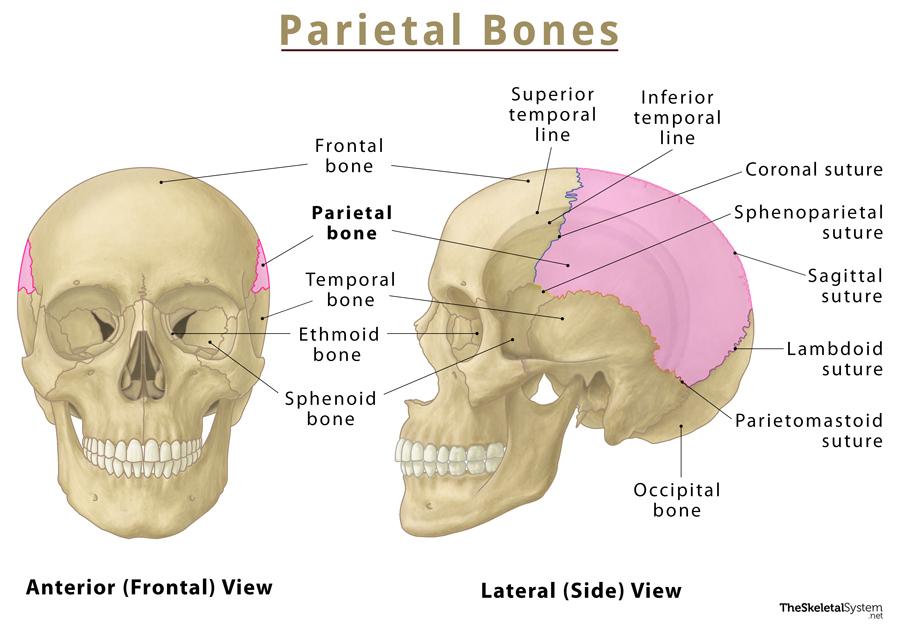
The temporal, one of the eight cranial bones, is a paired bone that forms a significant portion of the base and lateral wall of the skull. They protect the temporal lobe of the brain, hence the name.
Where is the Temporal Bone Located
The temporal bones are located on the lateral portions of the cranial floor.
Quick Facts
| Type | Irregular bone |
| How many are there in the human body | 2 (1 on each side of the skull) |
| Articulates with | 7 skull bones: Sphenoid, parietal, occipital, zygomatic, mandible, frontal, and maxilla |
Functions
- Provide structural support to the skull.
- Protect the temporal lobe of the brain.
- Help in the opening and closing of the mouth, in association with the mandible.
- Surround the ear canal and protect auditory nerves and internal ear structures that play a role in controlling hearing and balance.
Temporal Bone Anatomy
The temporal bone can be divided into three main parts: squamous, petromastoid, and tympanic.
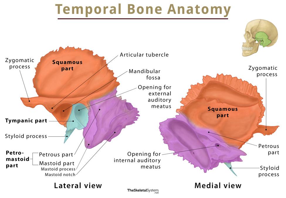
1. Squamous Part
Also called the squama temporalis, it is the largest part of the temporal bone, located anterosuperiorly. This flat and plate-like part forms the lateral part of the middle cranial fossa. Its outer surface is smooth and slightly convex and forms a part of the temporal fossa. On the other hand, its inner surface is concave, bearing impressions of the temporal lobe, specifically the folds (sulci and gyri). The inner surface also has a groove through which the middle meningeal vessels pass.
The lower portion of the squamous part bears an anterior bony projection called the zygomatic process. In the beginning, the process projects laterally, then it changes direction and travels anteriorly to articulate with the temporal process of the zygomatic bone, forming the zygomatic arch. At the zygomatic process’s root is a small tubercle called the articular tubercle or articular process. This tubercle forms the anterior boundary of the mandibular fossa, a socket-like structure through which the bone articulates with the mandible, forming the temporomandibular joint. This part of the bone also articulates with the sphenoid and parietal bone, anteriorly and laterally, respectively.
2. Petromastoid Part
It is composed of two parts: the mastoid, and petrous.
i. Mastoid Part
It is the posteriormost part of the temporal bone. Its outer surface is roughened by muscular attachments. This part bears an inferior conical bony projection called the mastoid process, which is palpable just behind the ear. On the medial surface of the mastoid process, there is a depression called the mastoid notch, and a deep groove called the sigmoid sulcus. The sigmoid sinus is located in this sigmoid sulcus. This part also contains mastoid air cells, some hollowed-out areas within the temporal bone. These cells act as a reservoir of air, protect the delicate structures of the ear, and equalize the air pressure within the middle ear.
ii. Petrous Part
It is a pyramid-shaped portion of the bone that projects medially and anteriorly from the squamous part. It forms a bony mass between the sphenoid and occipital bones within the cranial cavity. This part divides the middle and posterior cranial fossae. Its internal surface is grooved by the inferior temporal gyrus and trigeminal ganglion. The petrous part laterally articulates with the squamous part of the temporal bone, while the posterior area is continuous with the inner surface of the mastoid part.
3. Tympanic Part
Lying inferiorly to the squamous and anteriorly to the petromastoid part, it is a curved plate-like part located just below the zygomatic process. It fuses with the petrous part internally and the squamous and mastoid parts posteriorly. The concave posterior surface forms the anterior wall, floor, and part of the posterior wall of the external auditory canal. The external opening of the auditory canal is visible here. A narrow bony projection, called the styloid process, extends downwards and anteriorly from the inferior surface of the tympanic part. It is usually straight, but can sometimes have a curvature, usually on the anterior surface. A foramen, named stylomastoid foramen, lies between the styloid process and the mastoid process.
Articulations
As stated, the temporal bone articulates with seven skull bones, sphenoid, parietal, occipital, zygomatic, mandible, frontal, and maxilla. Among these, it only articulates with the mandible via a synovial joint called temporomandibular joint (TMJ). It gets fused with the remaining bones via respective sutures.
Muscle Attachments
Muscles that attach to the temporal bone are as follows:
- Temporalis muscle
- Sternocleidomastoid
- Splenius capitis
- Longissimus capitis
- Digastrics
- Stylopharyngeus
- Styloglossus
- Stylohyoid muscles
Ossification and Development
The temporal bone is ossified from eight centers: one for the squamous part, four for the petrous and mastoid parts, one for the tympanic part, and two for the styloid process.
References
- Temporal bone – Kenhub.com
- The Temporal Bone – Teachmeanatomy.info
- Temporal bone – Radiopaedia.org
- Temporal Bone Anatomy – Getbodysmart.com
- Temporal bone – Anatomy.app