Distal Phalanx
What is the Distal Phalanx
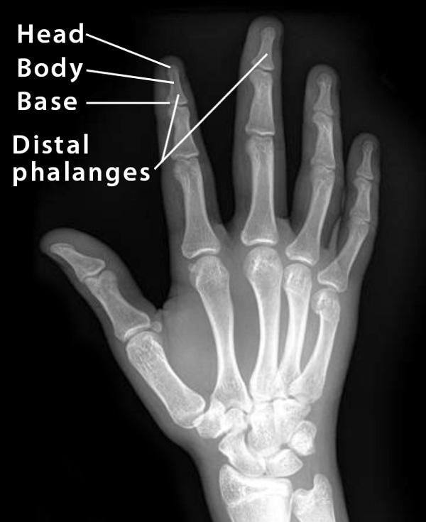
A distal phalanx is one of the tubular long bones found in each of the fingers [1, 2]. It is also referred to as the terminal phalanx. Like the other long bones in the hands, each distal phalanx is separated into a head, body or shaft, and a base [3].
Where are the Distal Phalanges Located
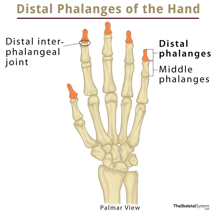
As suggested by the name, these phalanges form the distal row of finger bones, with each phalanx located distally to the middle phalanx in each finger. In the thumb, however, the distal phalanx in located directly above the proximal phalanx as there is no middle phalanx present here [4].
Distal Phalanx
How Many Distal Phalanges are There in the Fingers
There is one distal phalanx in each finger, including the thumb, which makes 5 in total for each hand, and 10 for both hands. These may be specified as the first (thumb), second (index finger), third (middle finger), fourth (ring finger), and fifth (little finger) distal phalanx.[/ps2id_wrap]
Development and Ossification
There is one primary ossification center for the body of each distal phalanx, appearing as early as the 8th-9th week of fetal growth. The secondary ossification center is for the base, and it develops when a child is 4-5 years old. These centers unite around the age of 18-20 years when the ossification of the phalanges is complete [5].
Distal Phalanges of the Hand X-Ray Image
Anatomy of the Distal Phalanges
Surfaces and Joints
Distal Interphalangeal (DIP) Joints (between the distal and middle phalanges in each finger) — The base or proximal end of the distal phalanges has a wide facet for articulating with the head or distal end of the middle phalanges in each finger [6]. In case of the thumb, this is the only Interphalangeal joint, formed between the distal and proximal phalanges [7]. You can find the DIP joint in the fingers, just below the fingernails.
The distal end or head has a tapered appearance, with a rough surface. On the dorsal surface, there is a spade-shaped rough plate, called the apical tuft or tuberosity of the distal phalanx, facing the palmar side [6].
Muscular Attachments
The head area is especially rough on the palmar surface as the tendons of the flexor digitorum profundus (FDP) muscle inserts here (in all the fingers, except the thumb) [8].
The thumb gets its terminal extensor tendon attachment from the extensor pollicis longus (EPL) muscle. The radial collateral ligament (RCL), and ulnar collateral ligament (UCL) are also vital in the stability and proper functioning of the thumb [7].
Blood Supply
Branches from the superficial palmar arch and deep palmar arch are responsible for blood supply in the distal phalangeal area [9].
Functions of the Distal Phalanges
The primary purpose of the distal phalanges is to support and maintain the stability of the fingertips, as it is a highly sensitive area with multiple nerve endings for translating the sense of touch into nerve impulses to be carried to the brain.
Additionally, the apical tufts or the horseshoe-shaped extensions of these bones support the fingernails on the back of the fingers, as well as the fleshy pads of the fingertips on the palmar side [10, 11].
Common Injuries and Associated Conditions
Fracture and Dislocation: The distal phalanges are among the most fractured bones of the hand, with those of the thumb and middle finger being affected most commonly, and the index finger being somewhat less frequently injured. Fractures and dislocations most often occur due to a direct blow to the fingers and may be associated with cuts and injuries to the fingertip or nail bed [12]. Mallet fractures and jersey fractures are among the common forms of fracture of these bones [13]. Treatment may include standard methods like splinting and reduced movement, along with proper care for any cuts or lesions.
Arthritis: Another common condition associated with this bone, the DIP joint of the index finger is more prone to arthritis due to its use in all pinching movements [7]. Standard treatment usually includes anti-inflammatory medicines, reduced movement, and splints, with surgeries in severe cases.
References
- http://anatomyzone.com/anatomy-feed/proximal-phalanx/
- http://library.open.oregonstate.edu/aandp/chapter/6-2-bone-classification/
- https://www.earthslab.com/anatomy/bones-of-the-hand-carpals-metacarpals-and-phalanges/
- https://radiopaedia.org/articles/phalanges-of-the-hands
- https://www.bartleby.com/107/56.html
- https://www.kenhub.com/en/library/anatomy/the-phalanges
- http://www.assh.org/handcare/Anatomy/Joints
- https://study.com/academy/lesson/flexor-digitorum-profundus-origin-action-insertion.html
- https://www.orthobullets.com/hand/6007/blood-supply-to-hand
- https://www.healthline.com/human-body-maps/distal-phalanges-hand
- https://www.imaios.com/en/e-Anatomy/Anatomical-Parts/Tuberosity-of-distal-phalanx
- https://www2.aofoundation.org/wps/portal/!ut/p/a0/04_Sj9CPykssy0xPLMnMz0vMAfGjzOKN_A0M3D2DDbz9_UMMDRyDXQ3dw9wMDAzMjfULsh0VAbWjLW0!/?bone=Hand&classification=75-Distal%20and%20Shaft%2C%20Transverse&segment=PhalangesDistal&showPage=diagnosis&teaserTitle=Distal%20%26%20shaft%2C%20transverse&contentUrl=srg/75/01-Diagnosis/ao_srg_diag_78_40.jsp
- https://emedicine.medscape.com/article/98322-overview