Trapezium Bone
Definition: What is the Trapezium Bone
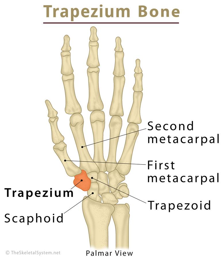
The trapezium (Latin: Os trapezium) is one of the eight carpal bones in the human hand [1], classified as a short bone like all other wrist bones [2]. The angularly shaped four-sided bone resembles and gets its name from the geometric shape trapezium [3]. Another name for the trapezium is the greater multangular bone [4].
Where is the Trapezium Bone Located
Located right at the base of the thumb, the trapezium is the first (or most lateral) bone in the distal carpal row [5]. It is situated between the first metacarpal, or the lowest bone in the thumb, and the scaphoid, with trapezoid at its side [6, 7].
Trapezium Bone
Development and Ossification
Like all the other carpal bones, the trapezium is cartilaginous at birth, beginning to ossify (becoming visible in an x-ray) when a child is 4 to 6 years old, around the same time as the scaphoid and trapezoid [8].
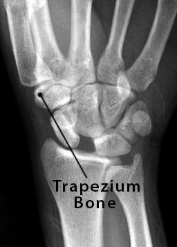
Trapezium X-Ray Image
Trapezium Bone Anatomy and Structure
Surfaces and Articulation
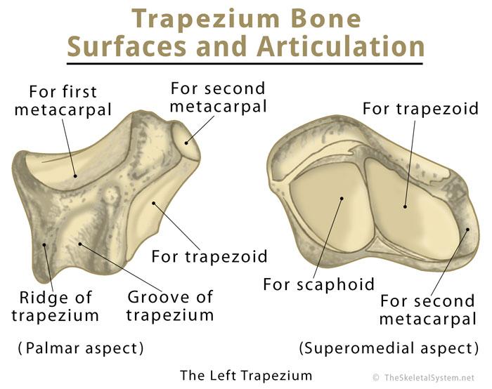
The trapezium articulates with two of the carpal bones, the scaphoid, and the trapezoid, as well as with the first metacarpal bone [7].
The proximal surface of the trapezium, in combination with the surface of the trapezoid, forms a slightly concave facet for articulating with the distal surface of the scaphoid [11].
The medial surface has a large concave facet that articulates with the trapezoid [7].
The distal surface forms a concave articular surface for the first metacarpal or the metacarpal of the thumb [9]. This forms the carpometacarpal joint of the thumb or trapeziometacarpal joint [10]. The trapezium may sometimes articulate with the metacarpal of the index finger (second metacarpal) as well, on the Inferomedial side [20].
Trapezium Bone Articular Surface Anatomy
The broad lateral surface of the bone is quite rough, mainly to accommodate ligament attachments, while the dorsal surface is smooth and does not have any articulation or attachments [7].
The palmar or volar surface has the primary muscular attachments [7], along with the groove of the trapezium [12], and the adjacent prominent bony ridge or tubercle [13].
Muscular Attachments
The muscles flexor pollicis brevis, opponens pollicis, and the abductor pollicis brevis arise from the above-mentioned tubercle [4].
Ligament Attachment
The radial collateral ligament is attached to the bone’s lateral surface [4], while the flexor retinaculum, the fibrous band that forms the roof of the carpal tunnel, attaches to both sides of the groove on the palmar surface [14].
Blood Supply
The radial artery provides primary blood supply through its distal branches, mainly via the bone’s non-articular dorsal surface [4]. Due to the rich and constant blood supply, the trapezium rarely develops the avascular necrosis [15], a condition common in some of the other carpal bones, occurring from inadequate blood circulation.
Functions: What Does the Trapezium Bone do
It plays an important role in shaping the distal joint of the wrist and maintaining its flexibility. The trapeziometacarpal or carpometacarpal joint of the thumb is most vital in the functioning of the hand. The only saddle joint in the human body [16], it allows the thumb to move more freely than any of the fingers, or the wrist, making it possible to grip objects for doing anything [10, 17].
Common Injuries and Associated Conditions
It is one of the less commonly fractured wrist bones, representing about 4% of all fractured carpal bones. A trapezium fracture usually occurs along with injury or fracture of other carpal and metacarpal bones, like in case of a serious accident [18].
Arthritis of the base of the thumb, referred to as the basilar thumb arthritis, develops when the cartilage in the trapezium-first metacarpal joint gets worn out. In severe cases, treatment may involve surgical removal of the trapezium [19].
References
- https://www.osmosis.org/learn/Trapezium_(bone)
- https://www.visiblebody.com/learn/skeleton/types-of-bones
- https://www.dummies.com/education/science/anatomy/bones-of-the-wrist-and-hand/
- https://radiopaedia.org/articles/trapezium
- https://www.ncbi.nlm.nih.gov/pubmedhealth/PMHT0023132/
- https://www.orthopaedicsone.com/display/Main/Trapezium
- https://www.healthline.com/human-body-maps/trapezium-bone
- http://sketchymedicine.com/2016/01/carpal-bone-ossification/
- http://www.mananatomy.com/body-systems/skeletal-system/carpal-bones-wrist
- https://www.jospt.org/doi/pdf/10.2519/jospt.2003.33.7.386
- https://classes.kumc.edu/sah/resources/handkines/bone/trapezium.html
- https://www.bartleby.com/107/pages/page225.html
- https://books.google.co.in/books?id=QFz-YOM0GCoC&pg=PA127&lpg=PA127&dq=trapezium+ridge+groove+tubercle&source=bl&ots=pUck5NwEZN&sig=pq_LtOrDUDkUescFbXVqJjoOtxc&hl=en&sa=X&ved=0ahUKEwimvvyG1ILcAhUEro8KHYuHDeEQ6AEIpQEwFQ#v=onepage&q=trapezium%20ridge%20groove%20tubercle&f=false
- https://www.sciencedirect.com/topics/medicine-and-dentistry/carpal-tunnel
- http://www.wheelessonline.com/ortho/trapezium
- https://www.knowyourbody.net/saddle-joint.html
- https://www.joionline.net/trending/content/trapezium-and-first-metacarpal-joint
- https://www.uptodate.com/contents/trapezium-fractures
- https://medicine.umich.edu/dept/orthopaedic-surgery/patient-care-services-hand-upper-extremity/basilar-thumb-arthritis
- https://www.kenhub.com/en/library/anatomy/carpal-bones