Zygomatic Bone (Cheekbone)
Published on February 18th 2022 by staff
What is the Zygomatic Bone
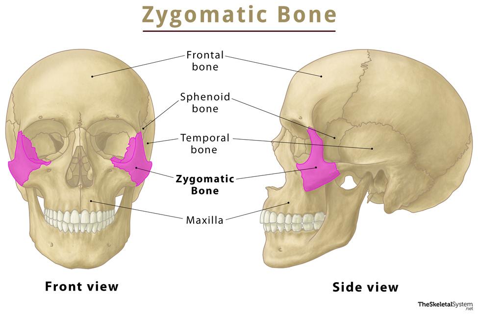
Zygomatic bone, commonly known as the cheekbone, is a paired, irregular facial bone. It is sometimes known as ‘zygoma’, a Greek word meaning ‘yoke’. This bone forms the cheeks and the lateral walls of the eye sockets or orbits.
Where is the Zygomatic Bone Located
It is located at the upper and lateral part of the face and can be felt from outside at the prominence of the cheekbones. More specifically, it is positioned just below each eye, extending upward to the outer side of each eye and downward near the jaw. You can easily feel the bone by touching the ridge above the fleshy area of the cheeks.
Quick Facts
| Type | Irregular bone |
| How many are there in the human body | 2, paired |
| Articulates with | Temporal bone, frontal bone, maxilla, and sphenoid bones. |
Functions
- It provides structure and strength to the mid-face, forming the cheek.
- The lower portion of the bone helps the otherwise immobile upper jawbone to do certain movements like speaking, chewing, drinking, coughing, and breathing by joining with it.
- It also protects the underneath arteries, nerves, veins, and organs.
Anatomy of the Zygomatic Bone
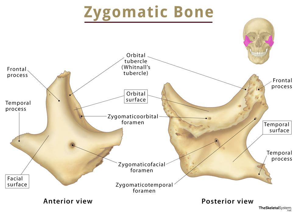
The bone is nearly quadrangular in shape, featuring three surfaces, five borders, and four processes.
Surfaces
The three surfaces of this bone are: facial, temporal, and orbital.
1. Facial or Malar Surface
Also known as the lateral surface as it faces outwards. It is smooth and convex, and has a small opening, the zygomaticofacial foramen, which allows passage to the zygomaticofacial nerve, vein, and artery from the face to the orbit. The zygomaticus major and the zygomaticus minor muscle attach to this surface’s anterior and posterior half.
2. Temporal Surface
This posteromedial surface of the bone is commonly called the temporal surface, as it faces towards the temporal bone and infratemporal fossae. The anterior region of this surface is rough and articulates with the zygomatic or malar process of the maxilla via the zygomaticomaxillary suture.
It includes the medial side of the bone’s temporal process, partially forming the lateral wall of the infratemporal fossa.
The temporal surface features the zygomaticotemporal foramen near the base of the frontal process, through which the zygomaticotemporal nerve transmits from the eye orbit to the temporal fossa.
3. Orbital Surface
This smooth concave surface faces the orbit, forming the major part of the lateral wall and half of the floor of the eye socket. It has a zygomatico-orbital foramen that leads to the zygomatic canal that divides into the zygomaticofacial and zygomaticotemporal canals, opening into the zygomaticofacial and zygomaticotemporal foramina, respectively.
Borders
1. Anterosuperior or Orbital Border: Smooth, concave, and present between the lateral and orbital surfaces of the bone. This border forms the inferolateral boundary of the eye orbit.
2. Anteroinferior or Maxillary Border: Articulates with the upper jawbone, maxilla on both sides via the zygomaticomaxillary suture. It is also the attachment point for the levator labii superioris muscle.
3. Posterosuperior or Temporal Border: A sinuous border with a convex top and concave bottom. It is continuous with the posterior border of the frontal process and the superior border of the zygomatic arch. The zygomaticotemporal foramen is also present on this surface. The temporal fascia also attaches here.
4. Posteroinferior Border: The rough border that lets the masseter muscle get attached to the bone.
5. Posteromedial Border: A serrated border where the zygomatic bone articulates with the greater wing of the sphenoid above (sphenozygomatic suture) and the orbital surface of the maxilla below. A small free surface called the posteromedial margin between the articular surfaces forms the lateral border of the inferior orbital fissure. This surface also serves as an attachment site for masseter muscle.
Processes
The four processes of this bone include:
1. Temporal Process of Zygomatic Bone : A backwardly directed bony projection, having an oblique, serrated end. It originates from the lower half of the bone, articulating with the zygomatic process of the temporal bone to form the zygomatic arch at the temporozygomatic suture.
2. Frontal Process of Zygomatic Bone: Another projection originating from the upper margin of the bone. It articulates with the frontal bone above and the greater wing of sphenoid posteriorly via the zygomaticofrontal, and sphenozygomatic sutures, respectively. The process terminates at the frontozygomatic suture.
There is a bony tubercle on the orbital surface of this process, called Whitnall’s tubercle. It serves as an attachment site for the suspensory ligament of the eyeball, the lateral palpebral ligament, and the levator palpebrae superioris muscle.
3. Maxillary Process of Zygomatic Bone: Located on the anterosuperior angle of the bone, its lower margin articulates with the maxilla.
4. Orbital Process of Zygomatic Bone: This projection forms the lateral wall of the eye orbit and also a part of its floor.
Articulations
As mentioned above, the zygomatic bone articulates with the maxilla, sphenoid, frontal, and temporal bones through the following sutures:
- Zygomaticomaxillary suture
- Zygomaticosphenoidal suture
- Zygomaticofrontal suture
- Zygomaticotemporal suture
All 4 articulations involving the zygomatic bone are collectively referred to as the zygomaticomaxillary complex (ZMC).
Muscle and Ligament Attachments
- Zygomaticus major and zygomaticus minor: These paired zygomaticus muscles attach to the lateral surface of the bone. They bring the upper lips upward and out, thus helping us to smile.
- Masseter: This powerful muscle originates at the zygomatic arch. From there, it spreads to the lower jaw, helping to close the jaw.
- Lateral palpebrae ligament (part of the levator palpebrae superioris): The ligament of levator palpebrae superioris, which elevates the upper eyelid, is attached to the Whitnall tubercle of the frontal process of the zygomatic bone.
- Levator labii superioris: It is a triangular band of muscle that stretches from under the eye sockets to the upper lip muscles. Its origin is at both the zygomatic bone’s maxillary process and the maxilla bone’s zygomatic process.
Ossification and Development
The bone ossifies around the eighth week of fetal life, which appears lateral and just beneath the orbit.
References
- Zygomatic bone – Kenhub.com
- Anatomy, Head and Neck, Zygomatic – Ncbi.nlm.nih.gov
- Zygomatic Bone Anatomy – Getbodysmart.com
- Zygoma – Radiopaedia.org
- Zygomatic Bone – Sciencedirect.com