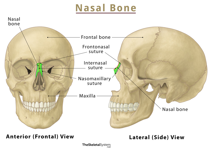
Sphenoid Bone
Published on April 18th 2022 by staff
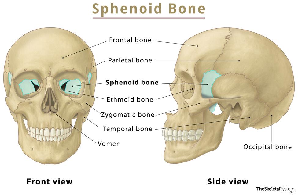
What is the Sphenoid Bone
The sphenoid is one of the eight cranial bones that make up the skull. It is a large, complex, unpaired bone, deriving its name from Greek’ sphenoeides’, meaning wedge-shaped. The bone forms a significant portion of the middle part of the skull base and the floor of the middle cranial fossa.
It is also called the “keystone” of the cranial floor since it is in contact with all other cranial bones.
Where is the Sphenoid Bone Located
It sits anteriorly in the cranium, just opposite the occipital bone.
Quick Facts
| Type | Irregular bone |
| How many are there in the human body | 1 |
| Articulates with | 12 bones: Parietal (paired), palatine (paired), zygomatic (paired), temporal (paired), frontal, ethmoid, occipital, and vomer |
Functions
It forms the base and lateral sides of the skull in combination with the orbital floor.
Anatomy: Parts and Structure of the Sphenoid Bone
The sphenoid bone consists of a central body, with two lateral paired wings on either side – the lesser and greater wings – and two pterygoid processes. This unique anatomy gives the bone a prominent bat-, butterfly-, or wasp-like appearance.
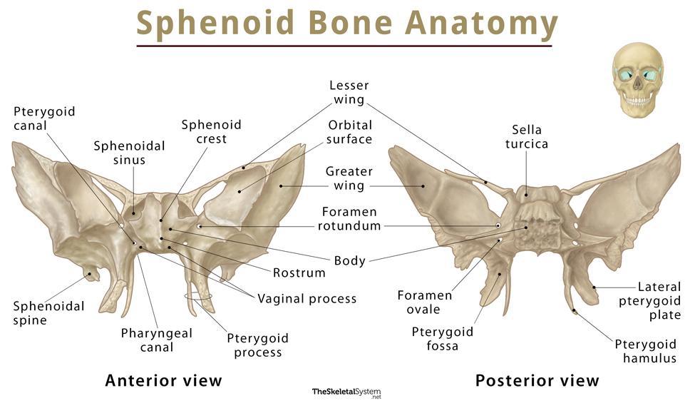
Body
As stated, the body is positioned centrally and is somewhat cubical. The body contains the sphenoidal sinuses, which are separated by a septum, opening into the nasal cavity. Due to the presence of these sinuses, the sphenoid body becomes a hollow structure.
Superior Surface
The superior surface of the sphenoid body contains the following bony landmarks:
1. Chiasmatic groove: It is a narrow transverse groove or sulcus formed by the optic chiasm that lies near the superior surface of the body. The groove ends on either side in the optic foramen, which transmits the optic nerve and blood vessels into the orbital cavity.
2. Sella turcica: It is a saddle-shaped depression having three parts:
i. Tuberculum sellae – This part forms the anterior wall of the sella turcica and the posterior aspect of the chiasmatic groove.
ii. Hypophyseal fossa – A small depression of the sella turcica, housing the pituitary gland.
iii. Dorsum sellae – It is a depression that slopes back at the base of the skull, forming the posterior wall of the sella turcica.
The sella turcica is surrounded by anterior and posterior clinoid processes that arise from the lesser wings and dorsum sellae. The tentorium cerebella muscle attaches here.
Inferior Surface
The inferior surface presents a triangular spine in the middle line, the sphenoidal spine or rostrum, which is continuous with the sphenoidal crest on the anterior surface. It also has a deep fissure between the alae of the vomer. On either side of the rostrum is a projecting lamina, called the vaginal process, located medially from the base of the medial pterygoid plate.
Lesser Wings
Two paired, triangular lesser wings arise from the front of the body of the sphenoid bone in a superolateral direction. It separates the anterior cranial fossa from the middle cranial fossa. These wings form the lateral border of the optic canal, through which the optic nerve and ophthalmic artery pass and reach the eye. The Superior surface of the lesser wings partially forms the cranial cavity, while their inferior surfaces contribute to the lateral margin of the bony orbit.
Greater Wings
The two greater wings emerge behind the lesser wings, extending in a lateral, superior, and posterior direction. They form the three parts of the facial skeleton: the floor of the middle cranial fossa, the skull’s lateral wall, and the orbit’s posterolateral wall.
The greater wings feature three foramina, which are as follows:
- Foramen rotundum: For the maxillary nerve
- Foramen ovale: Allows passage to the mandibular and lesser petrosal nerves, along with the accessory meningeal artery, and emissary vein
- Foramen spinosum: Conducts the middle meningeal vessels and a branch of the mandibular nerve
There is a slit or gap between the sphenoid body, lesser, and greater wings, known as the superior orbital fissure. This gap allows passage to several nerves and blood vessels of the bony orbit. These vessels include the trochlear, abducent, and oculomotor nerves, the ophthalmic nerve and its branches, and the superior ophthalmic vein.
Pterygoid Processes
The two pterygoid processes descend inferiorly from the point of junction between the sphenoid body and the greater wings. Each of these comprises a medial and a lateral pterygoid plate. The shorter and broader lateral plate is the point of origin for the medial and lateral pterygoid muscles. There is a small hook-like projection at the lower tip of the medial pterygoid plate, called the pterygoid hamulus.
The pterygoid and palatovaginal canals are also located in this process.
i. Pterygoid canal: It contains the major petrosal nerve and deep petrosal nerve.
ii. Palatovaginal or pharyngeal canal: It contains the pharyngeal nerve.
Muscle Attachments
- The temporalis muscle, a muscle of mastication, is attached to the temporal surface of the greater wing of the sphenoid.
- The upper fibers of the lateral pterygoid muscle attach to the infratemporal surface of the greater wing of the sphenoid as well as the infratemporal crest. This muscle also attaches to the lateral aspect of the lateral plate of the pterygoid process.
- The medial pterygoid muscle is attached to the medial aspect of the lateral plate as well as the pterygoid fossa.
- The medial division of the pterygoid process, called the medial plate, serves as a site of attachment to the superior pharyngeal constrictor muscle.
References
- The Sphenoid Bone – Teachmeanatomy.info
- Anatomy, Sphenoid Bone – Ncbi.nlm.nih.gov
- Sphenoid bone – Kenhub.com
- Sphenoid bone – Radiopaedia.org
- Sphenoid Bone – Sciencedirect.com