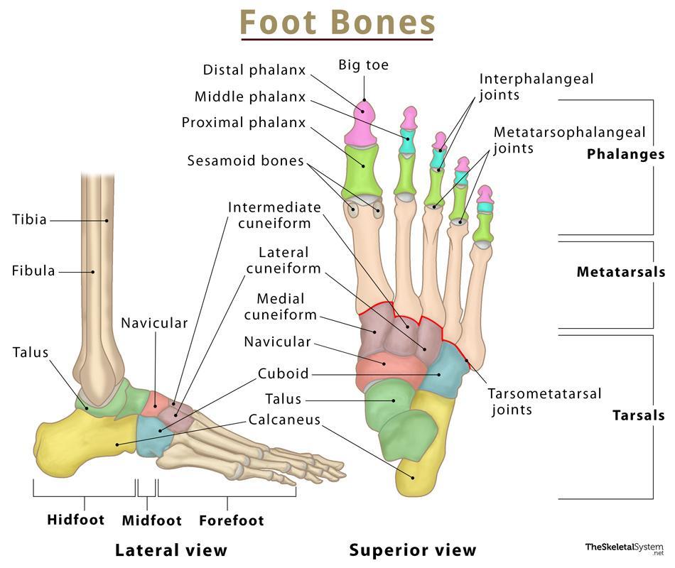
Foot Bones
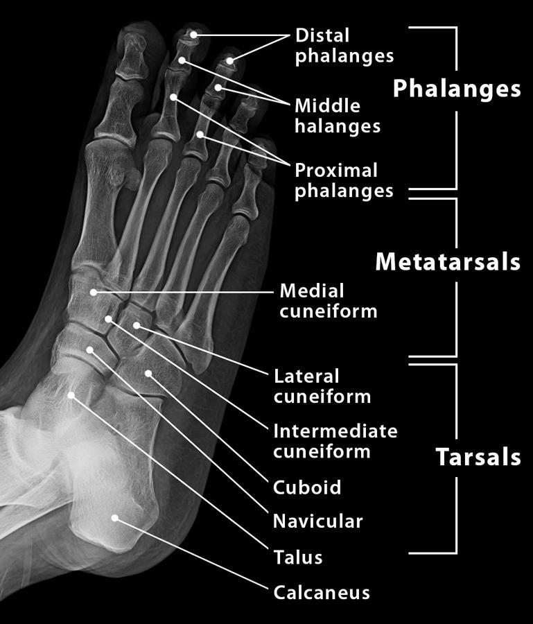
Humans have 26 bones in each foot that are classified into three groups – tarsals, metatarsals, and phalanges. These bones give structure to the foot and allow for all foot movements like flexing the toes and ankle, walking, and running.
The foot can be divided into three regions, the hindfoot, midfoot, and forefoot.
Names of the Bones in the Foot With Basic Anatomy
Tarsal Bones
The tarsals are a group of 7 irregular bones forming the hindfoot and the midfoot. These bones are arranged in two rows, proximal and distal. The bones in the proximal row form the hindfoot, while those in the distal row from the midfoot.
Hindfoot
The talus connects the foot to the rest of the leg and body through articulations with the tibia and fibula, the two long bones in the lower leg.
Midfoot
Some people may be born with an extra navicular bone (accessory navicular) beside the regular navicular bone, on the inside of the foot. This is a normal anatomical variation seen in around 2.5% of the entire population of the US.
Metatarsal Bones
These are a group of 5 long bones located towards the front of the foot, below the toes. These form the forefoot, along with the phalanges or toe bones.
These get shorter as we move from the big toe (hallux) towards the little toe and are numbered in that order.
Each of these bones has a head, body, and base. The base on their proximal side articulates with the carpal bones, while the head on the distal side articulates with the phalanges.
Phalanges
Also known as toe bones, these are the 14 long bones in the toes on each foot. As mentioned above, these form the forefoot with the metatarsals.
The second to fifth toes have 3 phalanges each, while only 2 are located in the big toe. These bones are named the proximal (closest to the ankle), middle and distal phalanges (farthest from the ankles) based on their location in the toes. The big toe only has the proximal and distal phalanx.
Being long bones, these are also anatomically divided into a head, body, and base.
There are two small ball-shaped sesamoid bones at the base of the big toe, near the joint between the 1st metatarsal and the proximal phalanx of the big toe. These bones act as the attachment point for multiple tendons and help with the movement of the big toe.
Joints Formed by the Foot Bones
In the Hindfoot
- Ankle joint: Synovial joint between talus, tibia, and fibula
- Subtalar joint: Between the talus and calcaneus
In the Midfoot
- Talonavicular: Between the talus and navicular bones
- Calcaneocuboid: Between the calcaneus and cuboid
- Intercunneiform: Among the three cuneiforms
- Tarsometatarsal (TMT): Between the distal tarsal bones and the base on the metatarsals
In the Forefoot
- Metatarsophalangeal (MTP): Between the head of the metatarsals and the base of the proximal phalanges.
- Interphalangeal joints: Between the phalanges on each toe. The big toe has only 1 interphalangeal joint, while the rest of the toes have 2 each.
Muscles, Ligaments, and Tendons
Many muscles, ligaments, and tendons are attached to the foot, which helps with all the small and large movements. The most important attachments are mentioned below.
Muscles
- Peroneal
- Anterior tibialis
- Posterior tibialis
- Extensors
- Flexors
Tendons and Ligaments
- Achilles tendon
- Plantar fascia
- Plantar calcaneonavicular ligament
- Calcaneocuboid ligament
References
- Foot bones: Everything you need to know – Medicalnewstoday.com
- Anatomy of the Foot and Ankle – Orthopaedia.com
- Foot Bones – Foot-pain-explored.com
- Anatomy of the Foot – Arthritis.org
- Feet (Human Anatomy): Bones, Tendons, Ligaments, and More – Webmd.com